|
|
|
|
|
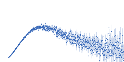
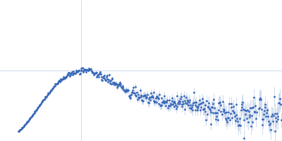
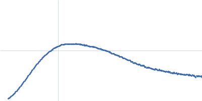
| Sample: |
Human IgG1 F(ab') 6C11 monomer, 24 kDa Homo sapiens protein
|
| Buffer: |
50 mM HEPES, 150 mM KCl, pH: 7.5 |
| Experiment: |
SAXS
data collected at BM29, ESRF on 2016 May 9
|
Antibody binding geometry and affinity control inhibitory FcγRIIB receptor signaling
Hayden Fisher
|
| RgGuinier |
2.5 |
nm |
| Dmax |
7.6 |
nm |
| VolumePorod |
56 |
nm3 |
|
|
|
|
|
|
|
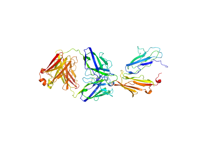
| Sample: |
Low affinity immunoglobulin gamma Fc region receptor II-b monomer, 20 kDa Homo sapiens protein
Human IgG1 F(ab') 6C11 monomer, 24 kDa Homo sapiens protein
|
| Buffer: |
50 mM HEPES, 150 mM KCl, pH: 7.5 |
| Experiment: |
SAXS
data collected at BM29, ESRF on 2016 May 9
|
Antibody binding geometry and affinity control inhibitory FcγRIIB receptor signaling
Hayden Fisher
|
| RgGuinier |
3.4 |
nm |
| Dmax |
12.0 |
nm |
| VolumePorod |
110 |
nm3 |
|
|
|
|
|
|
|
| Sample: |
Human IgG1 F(ab') 6G11 monomer, 25 kDa Homo sapiens protein
|
| Buffer: |
50 mM HEPES, 150 mM KCl, pH: 7.5 |
| Experiment: |
SAXS
data collected at BM29, ESRF on 2014 Dec 5
|
Antibody binding geometry and affinity control inhibitory FcγRIIB receptor signaling
Hayden Fisher
|
| RgGuinier |
2.6 |
nm |
| Dmax |
8.5 |
nm |
| VolumePorod |
60 |
nm3 |
|
|
|
|
|
|
|
| Sample: |
Low affinity immunoglobulin gamma Fc region receptor II-b monomer, 20 kDa Homo sapiens protein
Human IgG1 F(ab') 6G11 monomer, 25 kDa Homo sapiens protein
|
| Buffer: |
50 mM HEPES, 150 mM KCl, pH: 7.5 |
| Experiment: |
SAXS
data collected at BM29, ESRF on 2017 Apr 9
|
Antibody binding geometry and affinity control inhibitory FcγRIIB receptor signaling
Hayden Fisher
|
| RgGuinier |
3.5 |
nm |
| Dmax |
13.5 |
nm |
| VolumePorod |
97 |
nm3 |
|
|
|
|
|
|
|
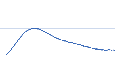
| Sample: |
Human IgG1 F(ab') 7C07 monomer, 25 kDa Homo sapiens protein
|
| Buffer: |
50 mM HEPES, 150 mM KCl, pH: 7.5 |
| Experiment: |
SAXS
data collected at BM29, ESRF on 2014 Dec 5
|
Antibody binding geometry and affinity control inhibitory FcγRIIB receptor signaling
Hayden Fisher
|
| RgGuinier |
2.6 |
nm |
| Dmax |
8.2 |
nm |
| VolumePorod |
59 |
nm3 |
|
|
|
|
|
|
|
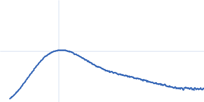
| Sample: |
Low affinity immunoglobulin gamma Fc region receptor II-b monomer, 20 kDa Homo sapiens protein
Human IgG1 F(ab') 7C07 monomer, 25 kDa Homo sapiens protein
|
| Buffer: |
50 mM HEPES, 150 mM KCl, pH: 7.5 |
| Experiment: |
SAXS
data collected at BM29, ESRF on 2014 Dec 5
|
Antibody binding geometry and affinity control inhibitory FcγRIIB receptor signaling
Hayden Fisher
|
| RgGuinier |
3.6 |
nm |
| Dmax |
13.5 |
nm |
| VolumePorod |
110 |
nm3 |
|
|
|
|
|
|
|
| Sample: |
Low affinity immunoglobulin gamma Fc region receptor II-b monomer, 20 kDa Homo sapiens protein
Human IgG1 F(ab') 6G08 monomer, 24 kDa Homo sapiens protein
|
| Buffer: |
50 mM HEPES, 150 mM KCl, pH: 7.5 |
| Experiment: |
SAXS
data collected at BM29, ESRF on 2016 Dec 14
|
Antibody binding geometry and affinity control inhibitory FcγRIIB receptor signaling
Hayden Fisher
|
| RgGuinier |
3.3 |
nm |
| Dmax |
12.0 |
nm |
| VolumePorod |
96 |
nm3 |
|
|
|
|
|
|
|
| Sample: |
H-NS family protein MvaT dimer, 12 kDa Pseudomonas putida (strain … protein
|
| Buffer: |
20 mM Tris-HCl, 500 mM NaCl, 10% glycerol, 500 mM Imidazole, pH: 8 |
| Experiment: |
SAXS
data collected at BL-10C, Photon Factory (PF), High Energy Accelerator Research Organization (KEK) on 2019 May 24
|
Structural characterization of the native oligomerization mode of MvaT proteins in Pseudomonas.
Microbiol Spectr :e0023526 (2026)
Vasileva D, Suzuki-Minakuchi C, Arakawa T, Moriwaki Y, Yonezawa K, Shimizu N, Fujimoto Z, Terada T, Okada K, Nojiri H
|
| RgGuinier |
2.9 |
nm |
| Dmax |
14.0 |
nm |
|
|
|
|
|
|
|
| Sample: |
Mutated ribosome assembly protein 1 (R1086Q) with C-terminal tag (RSRSGSENLYFQGSHHHHHHHH) monomer, 127 kDa Saccharomyces cerevisiae protein
|
| Buffer: |
50 mM HEPES, 300 mM NaCl, 5 mM MgCl2, 5% glycerol, pH: 8 |
| Experiment: |
SAXS
data collected at B21, Diamond Light Source on 2024 Oct 3
|
Hydroxyl radical footprinting modification reveals an intradomain communication pathway in EFL1 disrupted by a Shwachman-Diamond syndrome-associated mutation.
Protein Sci 35(4):e70504 (2026)
Zúñiga-Domínguez JA, Jain R, González-Andrade M, Farquhar ER, Chance MR, Gijsbers A, Sánchez-Puig N
|
| RgGuinier |
4.7 |
nm |
| Dmax |
15.2 |
nm |
| VolumePorod |
314 |
nm3 |
|
|
|
|
|
|
|
| Sample: |
Ribosome assembly protein 1 with C-terminal tag (RSRSGSENLYFQGSHHHHHHHH) monomer, 127 kDa Saccharomyces cerevisiae protein
|
| Buffer: |
50 mM HEPES, 300 mM NaCl, 5 mM MgCl2, 5% glycerol, pH: 8 |
| Experiment: |
SAXS
data collected at B21, Diamond Light Source on 2024 Oct 3
|
Hydroxyl radical footprinting modification reveals an intradomain communication pathway in EFL1 disrupted by a Shwachman-Diamond syndrome-associated mutation.
Protein Sci 35(4):e70504 (2026)
Zúñiga-Domínguez JA, Jain R, González-Andrade M, Farquhar ER, Chance MR, Gijsbers A, Sánchez-Puig N
|
| RgGuinier |
4.6 |
nm |
| Dmax |
15.7 |
nm |
| VolumePorod |
368 |
nm3 |
|
|
 6C11 experimental SAS data")
 6C11 experimental SAS data")
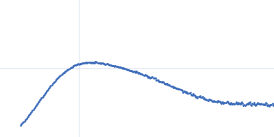
 6G11 experimental SAS data")
 6G11 experimental SAS data")
 7C07 experimental SAS data")
 7C07 experimental SAS data")
 6G08 experimental SAS data")

 with C-terminal tag (RSRSGSENLYFQGSHHHHHHHH) experimental SAS data")

 experimental SAS data")