|
|
|
|
|
| Sample: |
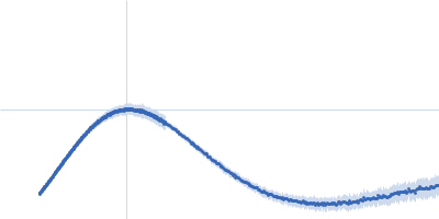
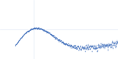
Ferric anguibactin-binding protein monomer, 32 kDa Bacillus cereus (strain … protein
|
| Buffer: |
20mM Tris-HCl, 100 mM NaCl, pH: 8 |
| Experiment: |
SAXS
data collected at 4C, Pohang Accelerator Laboratory on 2025 Apr 7
|
Structural basis of FatB-mediated iron uptake via tyrosine/histidine direct coordination accompanying long-distance domain reorganization.
Nat Commun (2026)
Lee H, Kim SO, You S, Segalina A, Noh T, Ihee H
|
| RgGuinier |
2.2 |
nm |
| Dmax |
7.4 |
nm |
| VolumePorod |
66 |
nm3 |
|
|
|
|
|
|
|
| Sample: |
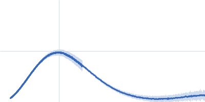
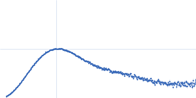
Ferric anguibactin-binding protein monomer, 32 kDa Bacillus cereus (strain … protein
|
| Buffer: |
20mM Tris-HCl, 100 mM NaCl, pH: 8 |
| Experiment: |
SAXS
data collected at 4C, Pohang Accelerator Laboratory on 2025 Apr 7
|
Structural basis of FatB-mediated iron uptake via tyrosine/histidine direct coordination accompanying long-distance domain reorganization.
Nat Commun (2026)
Lee H, Kim SO, You S, Segalina A, Noh T, Ihee H
|
| RgGuinier |
2.2 |
nm |
| Dmax |
7.5 |
nm |
| VolumePorod |
65 |
nm3 |
|
|
|
|
|
|
|
| Sample: |
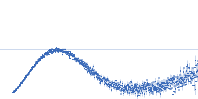
Ferric anguibactin-binding protein monomer, 32 kDa Bacillus cereus (strain … protein
|
| Buffer: |
20mM Tris-HCl, 100 mM NaCl, pH: 8 |
| Experiment: |
SAXS
data collected at 4C, Pohang Accelerator Laboratory on 2025 Apr 7
|
Structural basis of FatB-mediated iron uptake via tyrosine/histidine direct coordination accompanying long-distance domain reorganization.
Nat Commun (2026)
Lee H, Kim SO, You S, Segalina A, Noh T, Ihee H
|
| RgGuinier |
2.1 |
nm |
| Dmax |
7.1 |
nm |
| VolumePorod |
56 |
nm3 |
|
|
|
|
|
|
|
| Sample: |
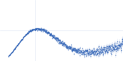
Ferric anguibactin-binding protein monomer, 32 kDa Bacillus cereus (strain … protein
|
| Buffer: |
20mM Tris-HCl, 100 mM NaCl, pH: 8 |
| Experiment: |
SAXS
data collected at 4C, Pohang Accelerator Laboratory on 2025 Apr 7
|
Structural basis of FatB-mediated iron uptake via tyrosine/histidine direct coordination accompanying long-distance domain reorganization.
Nat Commun (2026)
Lee H, Kim SO, You S, Segalina A, Noh T, Ihee H
|
| RgGuinier |
2.1 |
nm |
| Dmax |
7.1 |
nm |
| VolumePorod |
57 |
nm3 |
|
|
|
|
|
|
|
| Sample: |
Ferric anguibactin-binding protein monomer, 32 kDa Bacillus cereus (strain … protein
|
| Buffer: |
20mM Tris-HCl, 100 mM NaCl, pH: 8 |
| Experiment: |
SAXS
data collected at 4C, Pohang Accelerator Laboratory on 2025 Apr 7
|
Structural basis of FatB-mediated iron uptake via tyrosine/histidine direct coordination accompanying long-distance domain reorganization.
Nat Commun (2026)
Lee H, Kim SO, You S, Segalina A, Noh T, Ihee H
|
| RgGuinier |
2.1 |
nm |
| Dmax |
7.1 |
nm |
| VolumePorod |
42 |
nm3 |
|
|
|
|
|
|
|
| Sample: |
ABC transporter TM288 subunit monomer, 68 kDa Thermotoga maritima (strain … protein
ABC transporter, ATP-binding protein monomer, 64 kDa ABC transporter TM287 … protein
|
| Buffer: |
20 mM HEPES, 200 mM NaCl, 5 mM MgCl2, 20 μM DMM, 1 mM Mg2+ ATP, pH: 7.6 |
| Experiment: |
SAXS
data collected at EMBL P12, PETRA III on 2021 Sep 10
|
Capturing transient states of heterodimeric ABC transporter TM287/288 by Time-Resolved Small-Angle X-ray Scattering.
Biophys J (2026)
Schröder L, De Vecchis D, Gruzinov A, Schäfer LV, Blanchet CE, Seeger MA, Tidow H, Josts I
|
|
|
|
|
|
|
|
| Sample: |
ABC transporter TM288 subunit monomer, 68 kDa Thermotoga maritima (strain … protein
ABC transporter, ATP-binding protein monomer, 64 kDa ABC transporter TM287 … protein
Nanobody Nb_TM#1 bound to TM287/288 ABC transporter monomer, 14 kDa Vicugna pacos (alpaca)
|
| Buffer: |
20 mM HEPES, 200 mM NaCl, 5 mM MgCl2, 20 μM DMM, 1 mM Mg2+ ATP, pH: 7.6 |
| Experiment: |
SAXS
data collected at EMBL P12, PETRA III on 2021 Sep 10
|
Capturing transient states of heterodimeric ABC transporter TM287/288 by Time-Resolved Small-Angle X-ray Scattering.
Biophys J (2026)
Schröder L, De Vecchis D, Gruzinov A, Schäfer LV, Blanchet CE, Seeger MA, Tidow H, Josts I
|
|
|
|
|
|
|
|
| Sample: |
ABC transporter TM288 subunit monomer, 68 kDa Thermotoga maritima (strain … protein
ABC transporter, ATP-binding protein monomer, 64 kDa ABC transporter TM287 … protein
Synthetic nanobody Sb_TM#35 bound to TM287/288 ABC transporter monomer, 14 kDa synthetic (in vitro …
|
| Buffer: |
20 mM HEPES, 200 mM NaCl, 5 mM MgCl2, 20 μM DMM, 1 mM Mg2+ ATP, pH: 7.6 |
| Experiment: |
SAXS
data collected at EMBL P12, PETRA III on 2023 Jun 6
|
Capturing transient states of heterodimeric ABC transporter TM287/288 by Time-Resolved Small-Angle X-ray Scattering.
Biophys J (2026)
Schröder L, De Vecchis D, Gruzinov A, Schäfer LV, Blanchet CE, Seeger MA, Tidow H, Josts I
|
|
|
|
|
|
|
|
| Sample: |
Human IgG1 F(ab') 5C05 monomer, 46 kDa Homo sapiens protein
|
| Buffer: |
50 mM HEPES, 150 mM KCl, pH: 7.5 |
| Experiment: |
SAXS
data collected at BM29, ESRF on 2016 May 9
|
Antibody binding geometry and affinity control inhibitory FcγRIIB receptor signaling
Hayden Fisher
|
| RgGuinier |
2.5 |
nm |
| Dmax |
7.7 |
nm |
| VolumePorod |
59 |
nm3 |
|
|
|
|
|
|
|
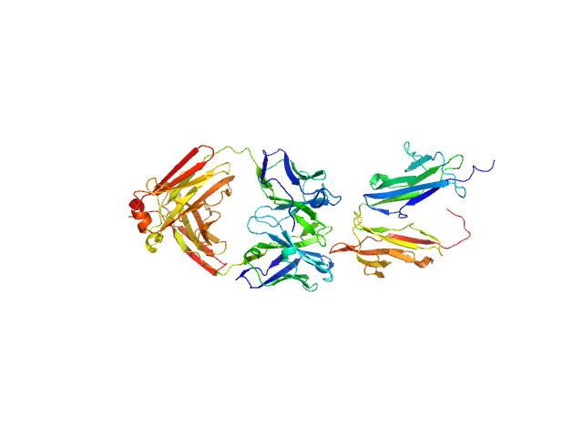
| Sample: |
Low affinity immunoglobulin gamma Fc region receptor II-b monomer, 20 kDa Homo sapiens protein
Human IgG1 F(ab') 5C05 monomer, 46 kDa Homo sapiens protein
|
| Buffer: |
50 mM HEPES, 150 mM KCl, pH: 7.5 |
| Experiment: |
SAXS
data collected at BM29, ESRF on 2016 May 18
|
Antibody binding geometry and affinity control inhibitory FcγRIIB receptor signaling
Hayden Fisher
|
| RgGuinier |
3.5 |
nm |
| Dmax |
14.0 |
nm |
| VolumePorod |
95 |
nm3 |
|
|

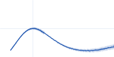

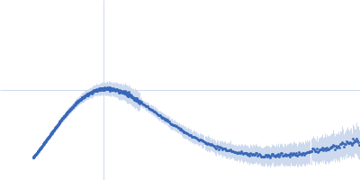

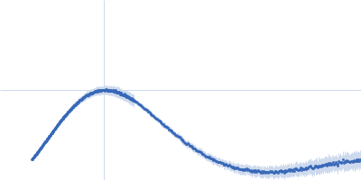


 5C05 experimental SAS data")
 5C05 experimental SAS data")