|
|
|
|
|
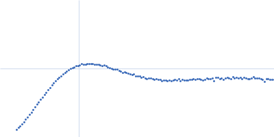
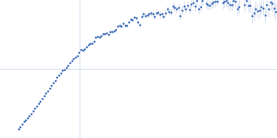
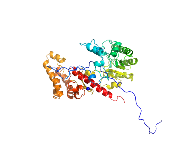
| Sample: |
Calmodulin-1 monomer, 17 kDa Homo sapiens protein
Pre-mRNA-processing factor 40 homolog A monomer, 9 kDa Homo sapiens protein
|
| Buffer: |
50 mM HEPES, 100 mM NaCl, 2 mM CaCl2, 1 mM TCEP, pH: 7.4 |
| Experiment: |
SAXS
data collected at 12.3.1 (SIBYLS), Advanced Light Source (ALS) on 2021 Sep 23
|
Binding by calmodulin is coupled to transient unfolding of the third FF domain of Prp40A.
Protein Sci 32(4):e4606 (2023)
Díaz Casas A, Cordoba JJ, Ferrer BJ, Balakrishnan S, Wurm JE, Pastrana-Ríos B, Chazin WJ
|
| RgGuinier |
2.4 |
nm |
| Dmax |
8.7 |
nm |
| VolumePorod |
29 |
nm3 |
|
|
|
|
|
|
|
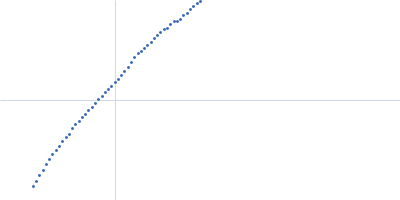
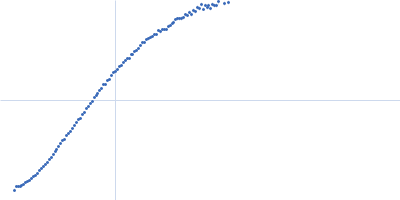
| Sample: |
Colibactin hybrid non-ribosomal peptide synthetase/type I polyketide synthase ClbK dimer, 476 kDa Escherichia coli protein
|
| Buffer: |
50 mM Hepes pH 7.5, 200 mM NaCl, 5 mM DTT, 5 mM MgCl2, pH: 7.5 |
| Experiment: |
SAXS
data collected at BM29, ESRF on 2020 Sep 14
|
Architecture of a PKS-NRPS hybrid megaenzyme involved in the biosynthesis of the genotoxin colibactin
Structure (2023)
Bonhomme S, Contreras-Martel C, Dessen A, Macheboeuf P
|
| RgGuinier |
8.0 |
nm |
| Dmax |
30.0 |
nm |
| VolumePorod |
1340 |
nm3 |
|
|
|
|
|
|
|
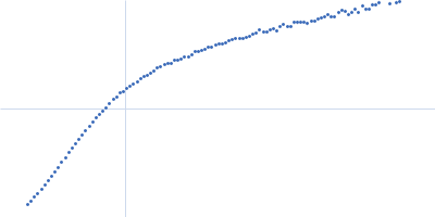
| Sample: |
Human dystrophin central domain R4-15 fragment monomer, 150 kDa protein
|
| Buffer: |
NaP 20 mM, NaCl 300 mM, EDTA 1 mM, Glycérol 2%, pH: 7.5 |
| Experiment: |
SAXS
data collected at SWING, SOLEIL on 2016 May 26
|
Dystrophin SAXS data
Raphael Dos Santos Morais
|
| RgGuinier |
9.7 |
nm |
| Dmax |
47.5 |
nm |
|
|
|
|
|
|
|
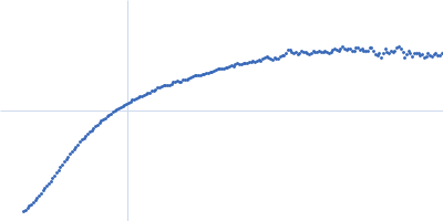
| Sample: |
Human dystrophin central domain R16-24 fragment monomer, 127 kDa protein
|
| Buffer: |
NaP 20 mM, NaCl 300 mM, EDTA 1 mM, Glycérol 2%, pH: 7.5 |
| Experiment: |
SAXS
data collected at SWING, SOLEIL on 2016 Dec 17
|
Dystrophin SAXS data
Raphael Dos Santos Morais
|
| RgGuinier |
9.3 |
nm |
| Dmax |
34.0 |
nm |
|
|
|
|
|
|
|
| Sample: |
Human dystrophin central domain R16-24 del45-55 fragment monomer, 58 kDa protein
|
| Buffer: |
NaP 20 mM, NaCl 300 mM, EDTA 1 mM, Glycérol 2%, pH: 7.5 |
| Experiment: |
SAXS
data collected at SWING, SOLEIL on 2017 Sep 28
|
Dystrophin SAXS data
Raphael Dos Santos Morais
|
| RgGuinier |
4.7 |
nm |
| Dmax |
18.3 |
nm |
|
|
|
|
|
|
|
| Sample: |
Human dystrophin central domain R16-24 del45-51 fragment monomer, 85 kDa protein
|
| Buffer: |
NaP 20 mM, NaCl 300 mM, EDTA 1 mM, Glycérol 2%, pH: 7.5 |
| Experiment: |
SAXS
data collected at SWING, SOLEIL on 2017 Jul 13
|
Dystrophin SAXS data
Raphael Dos Santos Morais
|
| RgGuinier |
7.0 |
nm |
| Dmax |
26.2 |
nm |
|
|
|
|
|
|
|
| Sample: |
Human dystrophin central domain R16-24 del44-54 fragment monomer, 57 kDa Homo sapiens protein
|
| Buffer: |
20 mM Na-phosphate, 300 mM NaCl, 1 mM EDTA, 2% glycerol,, pH: 7.5 |
| Experiment: |
SAXS
data collected at SWING, SOLEIL on 2019 Jul 8
|
Dystrophin SAXS data
Raphael Dos Santos Morais
|
| RgGuinier |
6.4 |
nm |
| Dmax |
25.0 |
nm |
| VolumePorod |
159 |
nm3 |
|
|
|
|
|
|
|
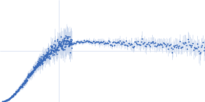
| Sample: |
Cell division protein FtsQ monomer, 11 kDa Mycobacterium tuberculosis (strain … protein
|
| Buffer: |
20 mM phosphate buffer, 25 mM NaCl, pH: 6.5 |
| Experiment: |
SAXS
data collected at 5-ID-D DND-CAT, Advanced Photon Source (APS), Argonne National Laboratory on 2017 Jun 28
|
An Arg/Ala-rich helix in the N-terminal region of M. tuberculosis FtsQ is a potential membrane anchor of the Z-ring.
Commun Biol 6(1):311 (2023)
Smrt ST, Escobar CA, Dey S, Cross TA, Zhou HX
|
| RgGuinier |
2.6 |
nm |
| Dmax |
8.5 |
nm |
| VolumePorod |
21 |
nm3 |
|
|
|
|
|
|
|
| Sample: |
DNA-guanine transglycosylase - D95A mutant monomer, 50 kDa Salmonella enterica subsp. … protein
|
| Buffer: |
100 mM KCl, 50 mM Tris pH 7.0, 1 mM DTT, pH: 7 |
| Experiment: |
SAXS
data collected at 12.3.1 (SIBYLS), Advanced Light Source (ALS) on 2021 Dec 22
|
7-Deazaguanines in DNA: functional and structural elucidation of a DNA modification system.
Nucleic Acids Res (2023)
Gedara SH, Wood E, Gustafson A, Liang C, Hung SH, Savage J, Phan P, Luthra A, de Crécy-Lagard V, Dedon P, Swairjo MA, Iwata-Reuyl D
|
| RgGuinier |
2.7 |
nm |
| Dmax |
10.1 |
nm |
| VolumePorod |
67 |
nm3 |
|
|
|
|
|
|
|
| Sample: |
DGQHR domain-containing protein dimer, 88 kDa Salmonella enterica subsp. … protein
|
| Buffer: |
100 mM KCl, 50 mM Tris pH 7.0, 1 mM DTT, pH: 7 |
| Experiment: |
SAXS
data collected at 12.3.1 (SIBYLS), Advanced Light Source (ALS) on 2021 Dec 22
|
7-Deazaguanines in DNA: functional and structural elucidation of a DNA modification system.
Nucleic Acids Res (2023)
Gedara SH, Wood E, Gustafson A, Liang C, Hung SH, Savage J, Phan P, Luthra A, de Crécy-Lagard V, Dedon P, Swairjo MA, Iwata-Reuyl D
|
| RgGuinier |
3.4 |
nm |
| Dmax |
13.5 |
nm |
| VolumePorod |
126 |
nm3 |
|
|