|
|
|
|
|
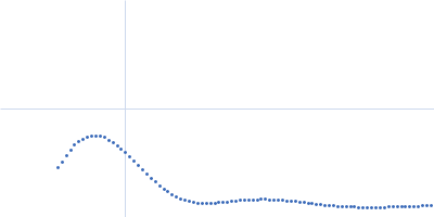
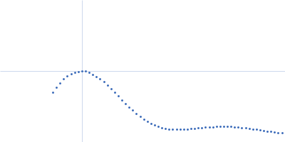
| Sample: |
CH848.3.D0949.10.17chim.6R.DS.SOSIP.664 Env glycoprotein trimer, 211 kDa HIV-1 group M protein
|
| Buffer: |
15 mM HEPES, 150 mM NaCl, pH: 7.1 |
| Experiment: |
SAXS
data collected at 14-ID-B (BioCARS), Advanced Photon Source (APS), Argonne National Laboratory on 2023 Mar 28
|
Microsecond dynamics control the HIV-1 Envelope conformation.
Sci Adv 10(5):eadj0396 (2024)
Bennett AL, Edwards R, Kosheleva I, Saunders C, Bililign Y, Williams A, Bubphamala P, Manosouri K, Anasti K, Saunders KO, Alam SM, Haynes BF, Acharya P, Henderson R
|
| RgGuinier |
4.1 |
nm |
| Dmax |
15.3 |
nm |
| VolumePorod |
768 |
nm3 |
|
|
|
|
|
|
|
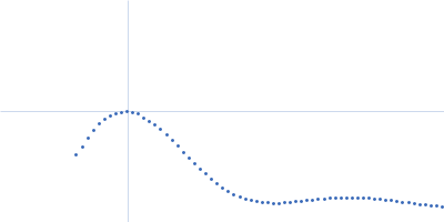
| Sample: |
CH848.3.D0949.10.17chim.6R.DS.SOSIP.664 Env glycoprotein trimer, 211 kDa HIV-1 group M protein
|
| Buffer: |
15 mM HEPES, 150 mM NaCl, pH: 7.1 |
| Experiment: |
SAXS
data collected at 14-ID-B (BioCARS), Advanced Photon Source (APS), Argonne National Laboratory on 2023 Mar 28
|
Microsecond dynamics control the HIV-1 Envelope conformation.
Sci Adv 10(5):eadj0396 (2024)
Bennett AL, Edwards R, Kosheleva I, Saunders C, Bililign Y, Williams A, Bubphamala P, Manosouri K, Anasti K, Saunders KO, Alam SM, Haynes BF, Acharya P, Henderson R
|
| RgGuinier |
5.3 |
nm |
| Dmax |
15.3 |
nm |
| VolumePorod |
765 |
nm3 |
|
|
|
|
|
|
|
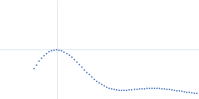
| Sample: |
CH848.3.D0949.10.17chim.6R.DS.SOSIP.664 Env glycoprotein trimer, 211 kDa HIV-1 group M protein
|
| Buffer: |
15 mM HEPES, 150 mM NaCl, pH: 7.1 |
| Experiment: |
SAXS
data collected at 14-ID-B (BioCARS), Advanced Photon Source (APS), Argonne National Laboratory on 2023 Mar 28
|
Microsecond dynamics control the HIV-1 Envelope conformation.
Sci Adv 10(5):eadj0396 (2024)
Bennett AL, Edwards R, Kosheleva I, Saunders C, Bililign Y, Williams A, Bubphamala P, Manosouri K, Anasti K, Saunders KO, Alam SM, Haynes BF, Acharya P, Henderson R
|
| RgGuinier |
5.3 |
nm |
| Dmax |
15.3 |
nm |
| VolumePorod |
764 |
nm3 |
|
|
|
|
|
|
|
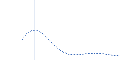
| Sample: |
CH848.3.D0949.10.17chim.6R.DS.SOSIP.664 Env glycoprotein trimer, 211 kDa HIV-1 group M protein
|
| Buffer: |
15 mM HEPES, 150 mM NaCl, pH: 7.1 |
| Experiment: |
SAXS
data collected at 14-ID-B (BioCARS), Advanced Photon Source (APS), Argonne National Laboratory on 2023 Mar 28
|
Microsecond dynamics control the HIV-1 Envelope conformation.
Sci Adv 10(5):eadj0396 (2024)
Bennett AL, Edwards R, Kosheleva I, Saunders C, Bililign Y, Williams A, Bubphamala P, Manosouri K, Anasti K, Saunders KO, Alam SM, Haynes BF, Acharya P, Henderson R
|
| RgGuinier |
5.2 |
nm |
| Dmax |
15.2 |
nm |
| VolumePorod |
728 |
nm3 |
|
|
|
|
|
|
|
| Sample: |
CH848.3.D0949.10.17chim.6R.DS.SOSIP.664 Env glycoprotein trimer, 211 kDa HIV-1 group M protein
|
| Buffer: |
15 mM HEPES, 150 mM NaCl, pH: 7.1 |
| Experiment: |
SAXS
data collected at 14-ID-B (BioCARS), Advanced Photon Source (APS), Argonne National Laboratory on 2023 Mar 30
|
Microsecond dynamics control the HIV-1 Envelope conformation.
Sci Adv 10(5):eadj0396 (2024)
Bennett AL, Edwards R, Kosheleva I, Saunders C, Bililign Y, Williams A, Bubphamala P, Manosouri K, Anasti K, Saunders KO, Alam SM, Haynes BF, Acharya P, Henderson R
|
| RgGuinier |
5.3 |
nm |
| Dmax |
15.1 |
nm |
| VolumePorod |
750 |
nm3 |
|
|
|
|
|
|
|
| Sample: |
CH848.3.D0949.10.17chim.6R.DS.SOSIP.664 Env glycoprotein trimer, 211 kDa HIV-1 group M protein
|
| Buffer: |
15 mM HEPES, 150 mM NaCl, pH: 7.1 |
| Experiment: |
SAXS
data collected at 14-ID-B (BioCARS), Advanced Photon Source (APS), Argonne National Laboratory on 2023 Mar 30
|
Microsecond dynamics control the HIV-1 Envelope conformation.
Sci Adv 10(5):eadj0396 (2024)
Bennett AL, Edwards R, Kosheleva I, Saunders C, Bililign Y, Williams A, Bubphamala P, Manosouri K, Anasti K, Saunders KO, Alam SM, Haynes BF, Acharya P, Henderson R
|
| RgGuinier |
5.3 |
nm |
| Dmax |
15.1 |
nm |
| VolumePorod |
752 |
nm3 |
|
|
|
|
|
|
|
| Sample: |
CH848.3.D0949.10.17chim.6R.DS.SOSIP.664 Env glycoprotein trimer, 211 kDa HIV-1 group M protein
|
| Buffer: |
15 mM HEPES, 150 mM NaCl, pH: 7.1 |
| Experiment: |
SAXS
data collected at 14-ID-B (BioCARS), Advanced Photon Source (APS), Argonne National Laboratory on 2023 Mar 30
|
Microsecond dynamics control the HIV-1 Envelope conformation.
Sci Adv 10(5):eadj0396 (2024)
Bennett AL, Edwards R, Kosheleva I, Saunders C, Bililign Y, Williams A, Bubphamala P, Manosouri K, Anasti K, Saunders KO, Alam SM, Haynes BF, Acharya P, Henderson R
|
| RgGuinier |
5.3 |
nm |
| Dmax |
15.1 |
nm |
| VolumePorod |
755 |
nm3 |
|
|
|
|
|
|
|
| Sample: |
CH848.3.D0949.10.17chim.6R.DS.SOSIP.664 Env glycoprotein trimer, 211 kDa HIV-1 group M protein
|
| Buffer: |
15 mM HEPES, 150 mM NaCl, pH: 7.1 |
| Experiment: |
SAXS
data collected at 14-ID-B (BioCARS), Advanced Photon Source (APS), Argonne National Laboratory on 2023 Mar 30
|
Microsecond dynamics control the HIV-1 Envelope conformation.
Sci Adv 10(5):eadj0396 (2024)
Bennett AL, Edwards R, Kosheleva I, Saunders C, Bililign Y, Williams A, Bubphamala P, Manosouri K, Anasti K, Saunders KO, Alam SM, Haynes BF, Acharya P, Henderson R
|
| RgGuinier |
5.3 |
nm |
| Dmax |
15.3 |
nm |
| VolumePorod |
741 |
nm3 |
|
|
|
|
|
|
|
| Sample: |
CH848.3.D0949.10.17chim.6R.DS.SOSIP.664 Env glycoprotein trimer, 211 kDa HIV-1 group M protein
|
| Buffer: |
15 mM HEPES, 150 mM NaCl, pH: 7.1 |
| Experiment: |
SAXS
data collected at 14-ID-B (BioCARS), Advanced Photon Source (APS), Argonne National Laboratory on 2023 Mar 30
|
Microsecond dynamics control the HIV-1 Envelope conformation.
Sci Adv 10(5):eadj0396 (2024)
Bennett AL, Edwards R, Kosheleva I, Saunders C, Bililign Y, Williams A, Bubphamala P, Manosouri K, Anasti K, Saunders KO, Alam SM, Haynes BF, Acharya P, Henderson R
|
| RgGuinier |
5.3 |
nm |
| Dmax |
15.1 |
nm |
| VolumePorod |
750 |
nm3 |
|
|