|
|
|
|
|
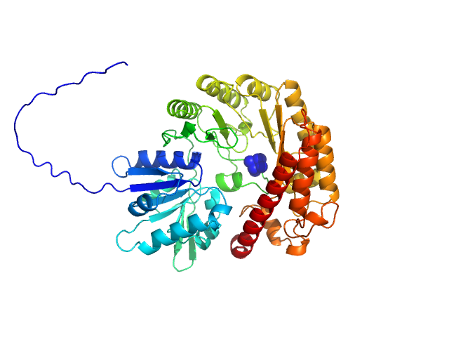
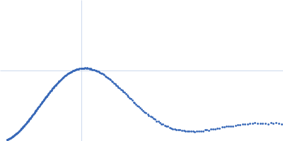
| Sample: |
Ubiquitin C-terminal hydrolase (UCH) domain of BAP1 wild type monomer, 27 kDa protein
|
| Buffer: |
20 mM HEPES, 150 mM NaCl, 3 mM CaCl2, 0.02% NaN3, pH: 7.5 |
| Experiment: |
SAXS
data collected at 13A, Taiwan Photon Source, NSRRC on 2024 Jun 23
|
Allosteric network of dynamic coupling within BAP1-UCH revealed by methyl NMR.
Nat Commun (2026)
Lai CH, Lou YC, Chang CF, Lu WL, Sriramoju MK, Wang YS, Wu KP, Camilloni C, Hsu SD
|
| RgGuinier |
2.1 |
nm |
| Dmax |
8.0 |
nm |
| VolumePorod |
35 |
nm3 |
|
|
|
|
|
|
|
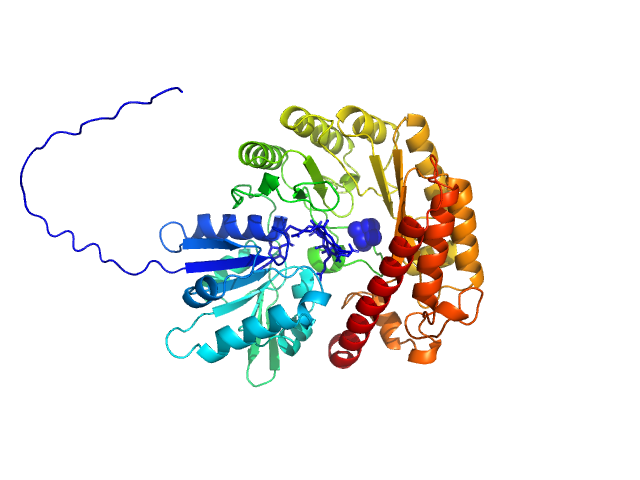
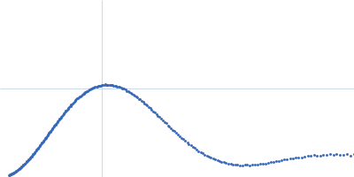
| Sample: |
Ubiquitin C-terminal hydrolase (UCH) domain of BAP1 L49V monomer, 27 kDa protein
|
| Buffer: |
20 mM HEPES, 150 mM NaCl, 1 mM TCEP, 0.02% NaN3, pH: 7.5 |
| Experiment: |
SAXS
data collected at 13A, Taiwan Photon Source, NSRRC on 2024 Jun 23
|
Allosteric network of dynamic coupling within BAP1-UCH revealed by methyl NMR.
Nat Commun (2026)
Lai CH, Lou YC, Chang CF, Lu WL, Sriramoju MK, Wang YS, Wu KP, Camilloni C, Hsu SD
|
| RgGuinier |
2.2 |
nm |
| Dmax |
8.7 |
nm |
| VolumePorod |
36 |
nm3 |
|
|
|
|
|
|
|
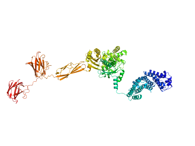
| Sample: |
B12-binding domain/radical SAM domain protein, MJ_0865 family monomer, 50 kDa Methanoculleus thermophilus protein
|
| Buffer: |
50 mM Tris, 300 mM NaCl, 5% glycerol, pH: 8 |
| Experiment: |
SAXS
data collected at SWING, SOLEIL on 2024 Sep 18
|
Insights into the stereochemical mechanism of the B(12)-dependent radical SAM glutamine C- methyltransferase (QCMT).
Commun Biol (2026)
Bourdin T, Guillot A, Mauger M, Lefranc B, Gervason S, Glousieau M, Grimaldi S, Leprince J, Thureau A, Benjdia A, Berteau O
|
| RgGuinier |
2.6 |
nm |
| Dmax |
10.4 |
nm |
| VolumePorod |
85 |
nm3 |
|
|
|
|
|
|
|
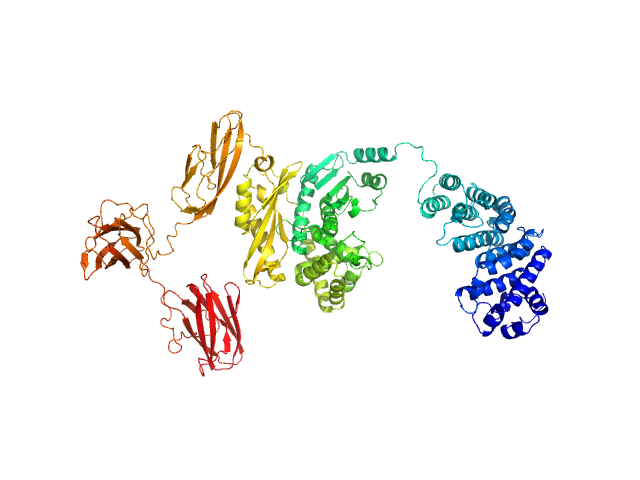
| Sample: |
B12-binding domain/radical SAM domain protein, MJ_0865 family monomer, 50 kDa Methanoculleus thermophilus protein
|
| Buffer: |
50 mM Tris, 300 mM NaCl, 5% glycerol, pH: 8 |
| Experiment: |
SAXS
data collected at SWING, SOLEIL on 2024 Sep 18
|
Insights into the stereochemical mechanism of the B(12)-dependent radical SAM glutamine C- methyltransferase (QCMT).
Commun Biol (2026)
Bourdin T, Guillot A, Mauger M, Lefranc B, Gervason S, Glousieau M, Grimaldi S, Leprince J, Thureau A, Benjdia A, Berteau O
|
| RgGuinier |
2.5 |
nm |
| Dmax |
10.1 |
nm |
| VolumePorod |
78 |
nm3 |
|
|
|
|
|
|
|
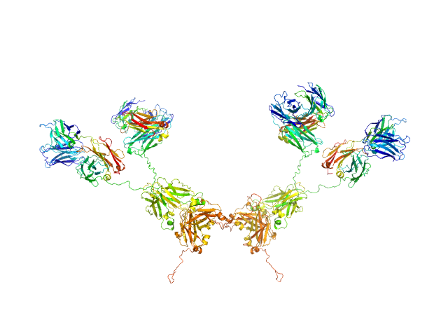
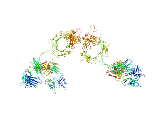
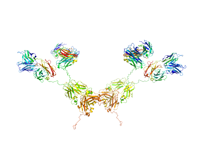
| Sample: |
Human immunoglobulin IgA1 dimer, 320 kDa protein
|
| Buffer: |
8.2 mM Na2HPO4, 1.5 mM KH2PO4, 137 mM NaCl, 2.6 mM KCl (PBS), pH: 7.4 |
| Experiment: |
SAXS
data collected at BM29, ESRF on 2014 Sep 7
|
Atomistic scattering modelling of the solution structure of human dimeric IgA1 reveals a structural and mechanistic basis for IgA nephropathy.
J Biol Chem :113156 (2026)
Bhatt JS, Yeo SC, Ireland SM, Ben-Younis A, Gor J, Molyneux K, Barratt J, Perkins SJ
|
| RgGuinier |
8.7 |
nm |
| Dmax |
35.0 |
nm |
|
|
|
|
|
|
|
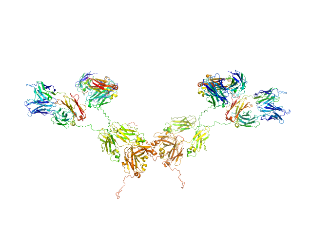
| Sample: |
Human immunoglobulin IgA1 dimer, 320 kDa protein
|
| Buffer: |
PBS buffer, 137 mM NaCl, 8.2 mM Na2HPO4, 2.6 mM KCl, 1.5 mM KH2PO4, pH: 7.4 |
| Experiment: |
SAXS
data collected at BM29, ESRF on 2014 Sep 7
|
Atomistic scattering modelling of the solution structure of human dimeric IgA1 reveals a structural and mechanistic basis for IgA nephropathy.
J Biol Chem :113156 (2026)
Bhatt JS, Yeo SC, Ireland SM, Ben-Younis A, Gor J, Molyneux K, Barratt J, Perkins SJ
|
| RgGuinier |
8.6 |
nm |
| Dmax |
35.0 |
nm |
|
|
|
|
|
|
|
| Sample: |
Human immunoglobulin IgA1 dimer, 320 kDa protein
|
| Buffer: |
PBS buffer, 137 mM NaCl, 8.2 mM Na2HPO4, 2.6 mM KCl, 1.5 mM KH2PO4, pH: 7.4 |
| Experiment: |
SAXS
data collected at BM29, ESRF on 2014 Sep 7
|
Atomistic scattering modelling of the solution structure of human dimeric IgA1 reveals a structural and mechanistic basis for IgA nephropathy.
J Biol Chem :113156 (2026)
Bhatt JS, Yeo SC, Ireland SM, Ben-Younis A, Gor J, Molyneux K, Barratt J, Perkins SJ
|
| RgGuinier |
8.4 |
nm |
| Dmax |
35.0 |
nm |
|
|
|
|
|
|
|
| Sample: |
Human immunoglobulin IgA1 dimer, 320 kDa protein
|
| Buffer: |
PBS buffer, 137 mM NaCl, 8.2 mM Na2HPO4, 2.6 mM KCl, 1.5 mM KH2PO4, pH: 7.4 |
| Experiment: |
SAXS
data collected at BM29, ESRF on 2014 Sep 7
|
Atomistic scattering modelling of the solution structure of human dimeric IgA1 reveals a structural and mechanistic basis for IgA nephropathy.
J Biol Chem :113156 (2026)
Bhatt JS, Yeo SC, Ireland SM, Ben-Younis A, Gor J, Molyneux K, Barratt J, Perkins SJ
|
| RgGuinier |
8.5 |
nm |
| Dmax |
35.0 |
nm |
|
|
|
|
|
|
|
| Sample: |
Collagenase ColG (E656D, N659T, G836V, D837Y) monomer, 114 kDa Hathewaya histolytica protein
|
| Buffer: |
10 mM HEPES, 100 mM NaCl, 1 mM CaCl2, pH: 7.5 |
| Experiment: |
SAXS
data collected at 12.3.1 (SIBYLS), Advanced Light Source (ALS) on 2022 Oct 28
|
Collagenase G
Cody Brazel
|
| RgGuinier |
4.7 |
nm |
| Dmax |
18.0 |
nm |
| VolumePorod |
181 |
nm3 |
|
|
|
|
|
|
|
| Sample: |
Collagenase ColG (E656D, N659T, G836V, D837Y) monomer, 114 kDa Hathewaya histolytica protein
|
| Buffer: |
10 mM HEPES, 100 mM NaCl, 1 mM CaCl2, EDTA, pH: 7.5 |
| Experiment: |
SAXS
data collected at 12.3.1 (SIBYLS), Advanced Light Source (ALS) on 2022 Oct 28
|
Collagenase G
Cody Brazel
|
| RgGuinier |
4.7 |
nm |
| Dmax |
19.0 |
nm |
| VolumePorod |
182 |
nm3 |
|
|
 domain of BAP1 wild type experimental SAS data")
 domain of BAP1 L49V experimental SAS data")


 experimental SAS data")
 experimental SAS data")