|
|
|
|
|
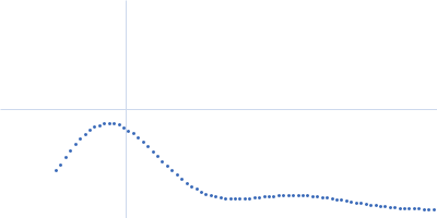
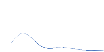
| Sample: |
CH505TFchim.6R.SOSIP.664 Env glycoprotein trimer, 217 kDa HIV-1 group M protein
|
| Buffer: |
15 mM HEPES, 150 mM NaCl, pH: 7.1 |
| Experiment: |
SAXS
data collected at 14-ID-B (BioCARS), Advanced Photon Source (APS), Argonne National Laboratory on 2022 Jul 30
|
Microsecond dynamics control the HIV-1 Envelope conformation.
Sci Adv 10(5):eadj0396 (2024)
Bennett AL, Edwards R, Kosheleva I, Saunders C, Bililign Y, Williams A, Bubphamala P, Manosouri K, Anasti K, Saunders KO, Alam SM, Haynes BF, Acharya P, Henderson R
|
| RgGuinier |
4.6 |
nm |
| Dmax |
15.5 |
nm |
| VolumePorod |
706 |
nm3 |
|
|
|
|
|
|
|
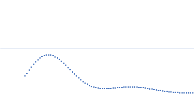
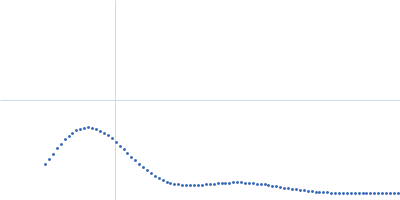
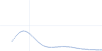
| Sample: |
CH505TFchim.6R.SOSIP.664 Env glycoprotein trimer, 217 kDa HIV-1 group M protein
|
| Buffer: |
15 mM HEPES, 150 mM NaCl, pH: 7.1 |
| Experiment: |
SAXS
data collected at 14-ID-B (BioCARS), Advanced Photon Source (APS), Argonne National Laboratory on 2022 Jul 28
|
Microsecond dynamics control the HIV-1 Envelope conformation.
Sci Adv 10(5):eadj0396 (2024)
Bennett AL, Edwards R, Kosheleva I, Saunders C, Bililign Y, Williams A, Bubphamala P, Manosouri K, Anasti K, Saunders KO, Alam SM, Haynes BF, Acharya P, Henderson R
|
| RgGuinier |
4.3 |
nm |
| Dmax |
15.3 |
nm |
| VolumePorod |
729 |
nm3 |
|
|
|
|
|
|
|
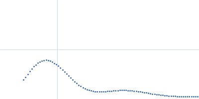
| Sample: |
CH505TFchim.6R.SOSIP.664 Env glycoprotein trimer, 217 kDa HIV-1 group M protein
|
| Buffer: |
15 mM HEPES, 150 mM NaCl, pH: 7.1 |
| Experiment: |
SAXS
data collected at 14-ID-B (BioCARS), Advanced Photon Source (APS), Argonne National Laboratory on 2022 Jul 28
|
Microsecond dynamics control the HIV-1 Envelope conformation.
Sci Adv 10(5):eadj0396 (2024)
Bennett AL, Edwards R, Kosheleva I, Saunders C, Bililign Y, Williams A, Bubphamala P, Manosouri K, Anasti K, Saunders KO, Alam SM, Haynes BF, Acharya P, Henderson R
|
| RgGuinier |
4.3 |
nm |
| Dmax |
15.4 |
nm |
| VolumePorod |
729 |
nm3 |
|
|
|
|
|
|
|
| Sample: |
CH505TFchim.6R.SOSIP.664 Env glycoprotein trimer, 217 kDa HIV-1 group M protein
|
| Buffer: |
15 mM HEPES, 150 mM NaCl, pH: 7.1 |
| Experiment: |
SAXS
data collected at 14-ID-B (BioCARS), Advanced Photon Source (APS), Argonne National Laboratory on 2022 Jul 28
|
Microsecond dynamics control the HIV-1 Envelope conformation.
Sci Adv 10(5):eadj0396 (2024)
Bennett AL, Edwards R, Kosheleva I, Saunders C, Bililign Y, Williams A, Bubphamala P, Manosouri K, Anasti K, Saunders KO, Alam SM, Haynes BF, Acharya P, Henderson R
|
| RgGuinier |
4.3 |
nm |
| Dmax |
15.3 |
nm |
| VolumePorod |
730 |
nm3 |
|
|
|
|
|
|
|
| Sample: |
CH505TFchim.6R.SOSIP.664 Env glycoprotein trimer, 217 kDa HIV-1 group M protein
|
| Buffer: |
15 mM HEPES, 150 mM NaCl, pH: 7.1 |
| Experiment: |
SAXS
data collected at 14-ID-B (BioCARS), Advanced Photon Source (APS), Argonne National Laboratory on 2022 Jul 28
|
Microsecond dynamics control the HIV-1 Envelope conformation.
Sci Adv 10(5):eadj0396 (2024)
Bennett AL, Edwards R, Kosheleva I, Saunders C, Bililign Y, Williams A, Bubphamala P, Manosouri K, Anasti K, Saunders KO, Alam SM, Haynes BF, Acharya P, Henderson R
|
| RgGuinier |
4.3 |
nm |
| Dmax |
15.3 |
nm |
| VolumePorod |
728 |
nm3 |
|
|
|
|
|
|
|
| Sample: |
CH505TFchim.6R.SOSIP.664 Env glycoprotein trimer, 217 kDa HIV-1 group M protein
|
| Buffer: |
15 mM HEPES, 150 mM NaCl, pH: 7.1 |
| Experiment: |
SAXS
data collected at 14-ID-B (BioCARS), Advanced Photon Source (APS), Argonne National Laboratory on 2022 Jul 28
|
Microsecond dynamics control the HIV-1 Envelope conformation.
Sci Adv 10(5):eadj0396 (2024)
Bennett AL, Edwards R, Kosheleva I, Saunders C, Bililign Y, Williams A, Bubphamala P, Manosouri K, Anasti K, Saunders KO, Alam SM, Haynes BF, Acharya P, Henderson R
|
| RgGuinier |
4.3 |
nm |
| Dmax |
15.3 |
nm |
| VolumePorod |
742 |
nm3 |
|
|
|
|
|
|
|
| Sample: |
CH505TFchim.6R.SOSIP.664 Env glycoprotein trimer, 217 kDa HIV-1 group M protein
|
| Buffer: |
15 mM HEPES, 150 mM NaCl, pH: 7.1 |
| Experiment: |
SAXS
data collected at 14-ID-B (BioCARS), Advanced Photon Source (APS), Argonne National Laboratory on 2022 Jul 29
|
Microsecond dynamics control the HIV-1 Envelope conformation.
Sci Adv 10(5):eadj0396 (2024)
Bennett AL, Edwards R, Kosheleva I, Saunders C, Bililign Y, Williams A, Bubphamala P, Manosouri K, Anasti K, Saunders KO, Alam SM, Haynes BF, Acharya P, Henderson R
|
| RgGuinier |
4.1 |
nm |
| Dmax |
15.4 |
nm |
| VolumePorod |
706 |
nm3 |
|
|
|
|
|
|
|
| Sample: |
CH505TFchim.6R.SOSIP.664 Env glycoprotein trimer, 217 kDa HIV-1 group M protein
|
| Buffer: |
15 mM HEPES, 150 mM NaCl, pH: 7.1 |
| Experiment: |
SAXS
data collected at 14-ID-B (BioCARS), Advanced Photon Source (APS), Argonne National Laboratory on 2022 Jul 29
|
Microsecond dynamics control the HIV-1 Envelope conformation.
Sci Adv 10(5):eadj0396 (2024)
Bennett AL, Edwards R, Kosheleva I, Saunders C, Bililign Y, Williams A, Bubphamala P, Manosouri K, Anasti K, Saunders KO, Alam SM, Haynes BF, Acharya P, Henderson R
|
| RgGuinier |
4.1 |
nm |
| Dmax |
15.5 |
nm |
| VolumePorod |
706 |
nm3 |
|
|