Tuukkanen A,
Freire D,
Chan S,
Arbing M,
Reed R,
Evans T,
Zenkeviciutė G,
Kim J,
Kahng S,
Sawaya M,
Chaton C,
Wilmanns M,
Eisenberg D,
Parret A,
Korotkov K,
(2018)
DOI
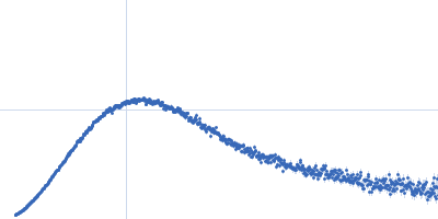
SASDDU2 – EspG3 chaperone from Mycobacterium smegmatis (Sel-Met labelled)
Synchrotron SAXS
data from solutions of
EspG3 chaperone from Mycobacterium smegmatis (Sel-Met labelled)
in
20 mM HEPES pH 7.5, 150 mM NaCl, pH 7.5
were collected
on the
EMBL P12 beam line
at the PETRA III storage ring
(DESY; Hamburg, Germany)
using a Pilatus 2M detector
at a wavelength of λ = 0.124 nm
(I(s) vs s, where s = 4πsinθ/λ, and 2θ is the scattering angle).
One solute concentration of 3.79 mg/ml was measured.
20 successive
0.050 second frames were collected.
The data were normalized to the intensity of the transmitted beam and radially averaged; the scattering of the solvent-blank was subtracted.
Cell temperature = UNKNOWN. Storage temperature = UNKNOWN. Sample detector distance = UNKNOWN
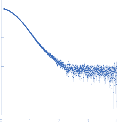 s, nm-1