Structure of ATP citrate lyase and the origin of citrate synthase in the Krebs cycle.

Verschueren KHG, Blanchet C Felix J, Dansercoer A, De Vos D, Bloch Y, Van Beeumen J, Svergun D, Gutsche I, Savvides SN, Verstraete K, Nature 568(7753):571-575 (2019) Europe PMC

SASDE56 – Human ATP-citrate synthase (ACLY) in HBS + Citrate + Coenzyme-A

ATP-citrate synthase
MWexperimental 480 kDa
MWexpected 458 kDa
VPorod 709 nm3
log I(s) 1.18×10⁴ 1.18×10³ 1.18×10² 1.18×101
ATP-citrate synthase small angle scattering data  s, nm-1
ln I(s)
ATP-citrate synthase Guinier plot ln 1.18×10⁴ Rg: 5.8 nm 0 (5.8 nm)-2 s2
(sRg)2I(s)/I(0)
ATP-citrate synthase Kratky plot 1.104 0 3 sRg
p(r)
ATP-citrate synthase pair distance distribution function Rg: 5.8 nm 0 Dmax: 16.5 nm

Data validation


Fits and models


log I(s)
 s, nm-1
ATP-citrate synthase SASREF CV model

log I(s)
 s, nm-1
ATP-citrate synthase MULTIFOXS model

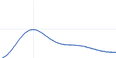
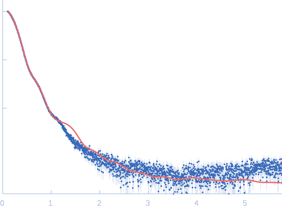
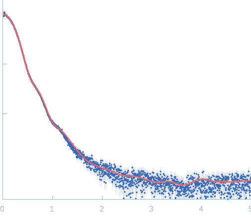
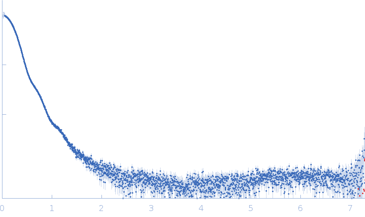
Synchrotron SAXS data from solutions of ATP-citrate synthase (ACLY) in 20mM HEPES, 150mM NaCl, 50mM Tris, 20mM citrate, 2mM coenzyme-A, pH 7.2 were collected on the EMBL P12 beam line at the PETRA III storage ring (Hamburg, Germany) using a Pilatus 6M detector at a sample-detector distance of 3.1 m and at a wavelength of λ = 0.124 nm (l(s) vs s, where s = 4πsinθ/λ, and 2θ is the scattering angle). Size-exclusion chromatography SAXS (SEC-SAXS) was employed using a sample injection concentration of 24.00 mg/ml. Data were measured at through the SEC elution at 20°C. 900 successive 1 second frames were collected. The data were normalized to the intensity of the transmitted beam and radially averaged; the scattering of an appropriate solvent-blank was subtracted using the CHROMIXS SEC-SAXS analysis package.

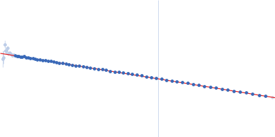
SEC column = UNKNOWN. Sample injection volume = UNKNOWN. Flow rate = UNKNOWN

ATP-citrate synthase
Mol. type   Protein
Organism   Homo sapiens
Olig. state   Tetramer
Mon. MW   114.6 kDa
 
UniProt   P53396-1
Sequence   FASTA