Structural Insights into Bacteriophage GIL01 gp7 Inhibition of Host LexA Repressor.

Caveney NA, Pavlin A, Caballero G, Bahun M, Hodnik V, de Castro L, Fornelos N, Butala M, Strynadka NCJ, Structure 27(7):1094-1102.e4 (2019) Europe PMC

SASDEC8 – Bacillus thuringiensis LexA repressor bound to Bacteriophage pGIL01 gp7 (Bt_LexA_GIL01_gp7)

Bacillus thuringiensis LexA repressor
Bacteriophage pGIL01 gp7
MWexperimental 108 kDa
MWexpected 70 kDa
log I(s) 1.53×100 1.53×10⁻¹ 1.53×10⁻² 1.53×10⁻³
Bacillus thuringiensis LexA repressor Bacteriophage pGIL01 gp7 small angle scattering data  s, nm-1
ln I(s)
Bacillus thuringiensis LexA repressor Bacteriophage pGIL01 gp7 Guinier plot ln 1.53×100 Rg: 4.4 nm 0 (4.4 nm)-2 s2
(sRg)2I(s)/I(0)
Bacillus thuringiensis LexA repressor Bacteriophage pGIL01 gp7 Kratky plot 1.104 0 3 sRg

Data validation


Fits and models


log I(s)
 s, nm-1
Bacillus thuringiensis LexA repressor Bacteriophage pGIL01 gp7 CHIMERA model

log I(s)
 s, nm-1
Bacillus thuringiensis LexA repressor Bacteriophage pGIL01 gp7 CHIMERA model

log I(s)
 s, nm-1
Bacillus thuringiensis LexA repressor Bacteriophage pGIL01 gp7 CHIMERA model

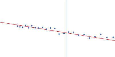
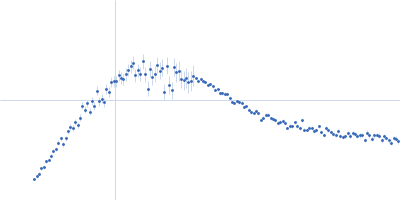
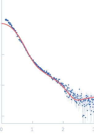
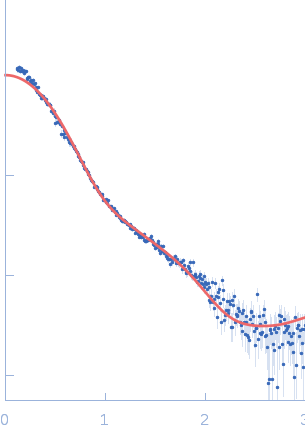
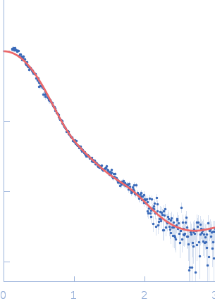
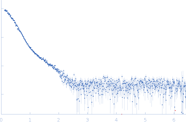
SAXS data from solutions of Bacillus thuringiensis LexA repressor bound to Bacteriophage pGIL01 gp7 (Bt_LexA_GIL01_gp7) in 20 mM HEPES, 300 mM NaCl, 10% glycerol, pH 8 were collected on a Rigaku BioSAXS-2000 instrument at the University of British Columbia (Vancouver, Canada) using a Hybrid Pixel Array Detector Rigaku HyOix-3000 detector at a wavelength of λ = 0.154 nm (l(s) vs s, where s = 4πsinθ/λ, and 2θ is the scattering angle). Solute concentrations ranging between 0.8 and 12 mg/ml were measured at 6°C. 12 successive 300 second frames were collected. The data were normalized to the intensity of the transmitted beam and radially averaged; the scattering of the solvent-blank was subtracted. The low angle data collected at lower concentration were merged with the highest concentration high angle data to yield the final composite scattering curve.

Sample detector distance = UNKNOWN

Bacillus thuringiensis LexA repressor (LexA)
Mol. type   Protein
Organism   Bacillus thuringiensis
Olig. state   Dimer
Mon. MW   23.4 kDa
 
UniProt   A0A2B4EEI3 (1-210)
Sequence   FASTA
 
Bacteriophage pGIL01 gp7 (gp7)
Mol. type   Protein
Organism   Bacteriophage pGIL01
Olig. state   Tetramer
Mon. MW   5.9 kDa
 
UniProt   Q7WSG2 (1-50)
Sequence   FASTA