Verschueren KHG,
Blanchet C
Felix J,
Dansercoer A,
De Vos D,
Bloch Y,
Van Beeumen J,
Svergun D,
Gutsche I,
Savvides SN,
Verstraete K,
Nature
568(7753):571-575
(2019)
Europe PMC
SASDFB3 – Human ATP-citrate synthers (ACLY) full length in HBS + Citrate
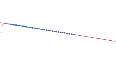
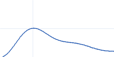
Synchrotron SAXS data from solutions of Human ATP-citrate synthers (ACLY) in 20mM HEPES, 150mM NaCl, 50mM Tris, 20mM citrate, pH 7.2 were collected on the EMBL P12 beam line at the PETRA III storage ring (Hamburg, Germany) using a Pilatus 2M detector at a sample-detector distance of 3.1 m and at a wavelength of λ = 0.124 nm (l(s) vs s, where s = 4πsinθ/λ, and 2θ is the scattering angle). Size-exclusion chromatography SAXS (SEC-SAXS) was employed using a sample injection concentration of 16.00 mg/ml. Data were measured at through the SEC elution at 20°C. 900 successive 1 second frames were collected. The data were normalized to the intensity of the transmitted beam and radially averaged; the scattering of an appropriate solvent-blank was subtracted using the CHROMIXS SEC-SAXS analysis package.
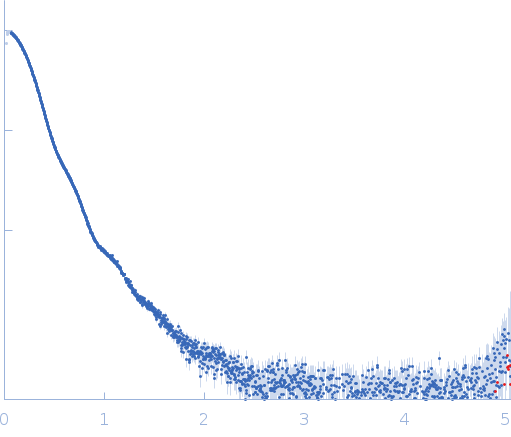 s, nm-1