A cocrystal structure of dengue capsid protein in complex of inhibitor.

Xia H, Xie X, Zou J, Noble CG, Russell WK, Holthauzen LMF, Choi KH, White MA, Shi PY, Proc Natl Acad Sci U S A 117(30):17992-18001 (2020) Europe PMC

SASDHJ4 – Dengue Virus 2 New Guinea C (DENV-2 C, capsid protein), apo form

Dengue Virus 2 New Guinea C
MWexperimental 21 kDa
MWexpected 19 kDa
VPorod 52 nm3
log I(s) 1.39×10⁴ 1.39×10³ 1.39×10² 1.39×101
Dengue Virus 2 New Guinea C small angle scattering data  s, nm-1
ln I(s)
Dengue Virus 2 New Guinea C Guinier plot ln 1.39×10⁴ Rg: 1.8 nm 0 (1.8 nm)-2 s2
(sRg)2I(s)/I(0)
Dengue Virus 2 New Guinea C Kratky plot 1.104 0 3 sRg
p(r)
Dengue Virus 2 New Guinea C pair distance distribution function Rg: 1.8 nm 0 Dmax: 5.4 nm

Data validation


Fits and models


log I(s)
 s, nm-1
Dengue Virus 2 New Guinea C CORAL model

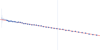
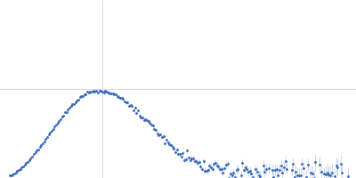
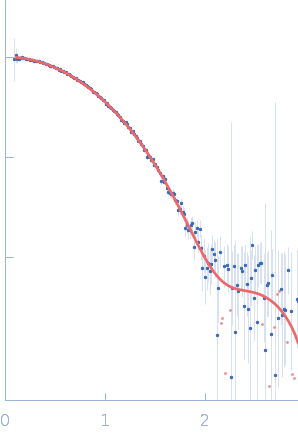
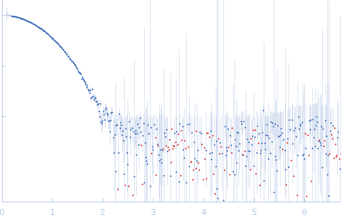
SAXS data from solutions of the DENV-2 C, capsid protein (apo form) in 100 mM NaCl, 25 mM HEPES, pH 7.4, pH 7.4 were collected using a Rigaku BioSAXS-1000 instrument (Sealy Center For Structural Biology, UTMB-G, Galveston, TX) equipped with a Pilatus 100K detector at a sample-detector distance of 0.5 m and at a wavelength of λ = 0.15418 nm (I(s) vs s, where s = 4πsinθ/λ, and 2θ is the scattering angle). One solute concentration of 2.50 mg/ml was measured at 10°C. 10 successive 3600 second frames were collected. The data were normalized to the intensity of the transmitted beam and radially averaged; the scattering of the solvent-blank was subtracted.

Dengue Virus 2 New Guinea C (DENV-2 C)
Mol. type   Protein
Organism   Dengue virus 2
Olig. state   Dimer
Mon. MW   9.4 kDa
 
UniProt   P14340 (21-100)
Sequence   FASTA