| MWI(0) | 24 | kDa |
| MWexpected | 25 | kDa |
|
log I(s)
1.90×10⁻³
1.90×10⁻⁴
1.90×10⁻⁵
1.90×10⁻⁶
|
 s, nm-1
|
|
|
|

|
|

|
|
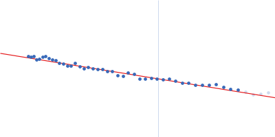
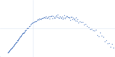
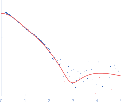
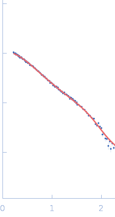
Synchrotron SAXS
data from solutions of
Binary-patterned 4-helix bundle de novo protein WA20
in
20 mM HEPES, 100 mM NaCl, 200 mM ArgHCl, 10% glycerol,, pH 7.5
were collected
on the
BL-6A beam line
at the Photon Factory (PF), High Energy Accelerator Research Organization (KEK) storage ring
(Tsukuba, Japan)
using a Pilatus3 1M detector
at a sample-detector distance of 1 m and
at a wavelength of λ = 0.15 nm
(I(s) vs s, where s = 4πsinθ/λ, and 2θ is the scattering angle).
One solute concentration of 5.00 mg/ml was measured
at 20°C.
30 successive
5 second frames were collected.
The data were normalized to the intensity of the transmitted beam and radially averaged; the scattering of the solvent-blank was subtracted.
Storage temperature = UNKNOWN |
|
|||||||||||||||||||||||||||