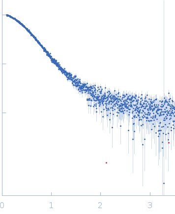
| MWI(0) | 33 | kDa |
| MWexpected | 29 | kDa |
| VPorod | 64 | nm3 |
|
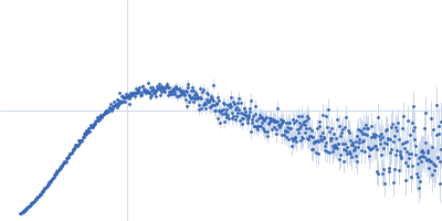
log I(s)
3.06×10³
3.06×10²
3.06×101
3.06×100
|
 s, nm-1
|
|
|
|

|
|
|
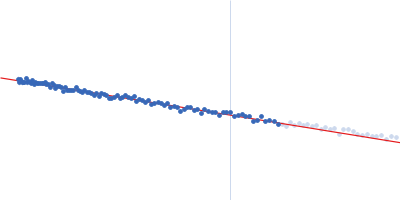
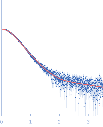
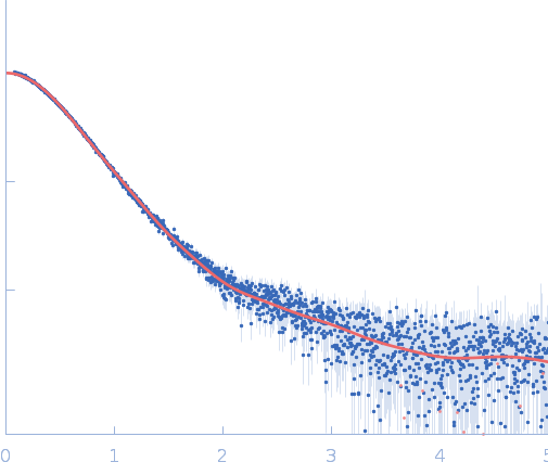
Synchrotron SAXS
data from solutions of
SARS-CoV-2 spike protein ACE2 receptor binding domain (RBD)
in
25 mM Tris 100 mM NaCl, pH 7.5
were collected
on the
EMBL P12 beam line
at the PETRA III storage ring
(DESY; Hamburg, Germany)
using a Pilatus 6M detector
at a sample-detector distance of 3 m and
at a wavelength of λ = 0.124 nm
(I(s) vs s, where s = 4πsinθ/λ, and 2θ is the scattering angle).
One solute concentration of 1.10 mg/ml was measured
at 20°C.
10 successive
0.100 second frames were collected.
The data were normalized to the intensity of the transmitted beam and radially averaged; the scattering of the solvent-blank was subtracted.
|
|
|||||||||||||||||||||||||||