|
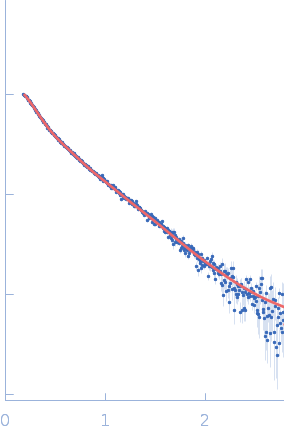
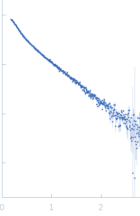
Synchrotron SAXS data from solutions of pri-miR16-1 primary microRNA in 50 mM KCl, 50 mM HEPES, 5 mM DTT, 1% glycerol, pH 7.5 were collected on the G1 beam line at the Cornell High Energy Synchrotron Source (CHESS; Ithaca, NY, USA) using a Pilatus 100k detector at a sample-detector distance of 1.5 m and at a wavelength of λ = 0.125 nm (I(s) vs s, where s = 4πsinθ/λ, and 2θ is the scattering angle). One solute concentration of 0.43 mg/ml was measured at 23°C. 20 successive 1 second frames were collected. The data were normalized to the intensity of the transmitted beam and radially averaged; the scattering of the solvent-blank was subtracted.
The bead models displayed in this entry show an individual reconstructed model (top) and associated individual model fit to the data and the volume-corrected and bead-occupancy representation obtained from the spatial alignment of several individual model reconstructions (DAMFILT; bottom).
|
|
 s, nm-1