|
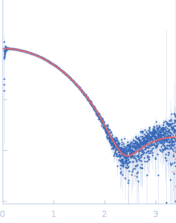
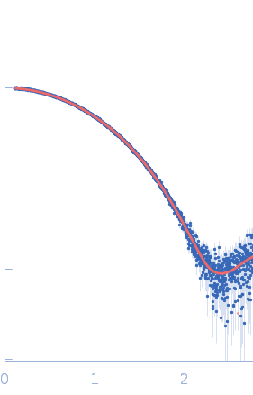
Synchrotron SAXS data from solutions of Wild-type human myelin protein P2 in 20 mM HEPES, 300 mM NaCl, 1 mM DTT, pH 7.5 were collected on the B21 beam line at the Diamond Light Source (Didcot, UK) using a Eiger 4M detector at a wavelength of λ = 0.095 nm (I(s) vs s, where s = 4πsinθ/λ, and 2θ is the scattering angle). In-line size-exclusion chromatography (SEC) SAS was employed. The SEC parameters were as follows: A sample at 9.9 mg/ml was injected at a 0.075 ml/min flow rate onto a GE Superdex 200 Increase 3.2/300 column at 20°C. 21 successive 3 second frames were collected through the SEC peak. The data were normalized to the intensity of the transmitted beam and radially averaged; the scattering of the solvent-blank was subtracted.
Storage temperature = UNKNOWN. Sample detector distance = UNKNOWN. Sample injection volume = UNKNOWN
|
|
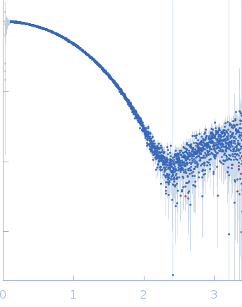 s, nm-1
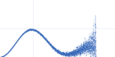