|
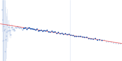
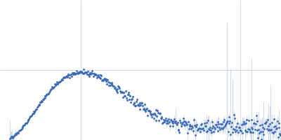
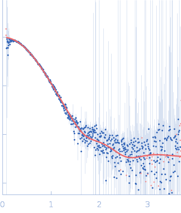
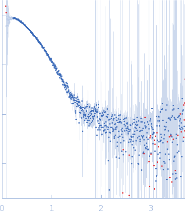
Synchrotron SAXS data from solutions of Arabidopsis thaliana beta-amylase 1 in 50 mM MES, 100 mM NaCl, 1 mM DTT, pH 6.7 were collected on the 12.3.1 (SIBYLS) beam line at the Advanced Light Source (ALS; Berkeley, CA, USA) using a Pilatus3 X 2M detector at a sample-detector distance of 1.5 m and at a wavelength of λ = 0.103 nm (I(s) vs s, where s = 4πsinθ/λ, and 2θ is the scattering angle).
BAM1 from Arabidopsis was expressed from a pET29a vector with the S-protein tag and C-terminal His tag. The plasmid of AtBAM1 was added to BL21 cell cultures. The cells containing the plasmid for AtBAM1 were then grown in 2xYT media (RPI) along with 1 μL mL−1 Kanamycin at 37 °C with shaking at 200 rpm. Once the growth reached an optical density of 0.6 at 600 nm, 0.75 mM of IPTG was added and the temperature was dropped to 20 oC. Cells shook overnight for about 10-12 hours, and then were pelleted by centrifugation at 5,000 rpm (3,024 rcf) at 4 oC for 15 minutes. Pellets were then frozen at -80oC until needed. Pellets were thawed and resuspended in a buffer composed of 50mM NaH2PO4, 0.5 M NaCl, and 2mM Imidazole (pH 5) with one protease inhibitor tablet (Pierce A32965). Cells were lysed by sonication in an ice bath for 3 minutes at 60% amplitude (5 seconds pulse on; 5 seconds pulse off). The cell lysate then underwent clarification by centrifugation at 15,000 rpm (27,216 rcf) at 4oC for 15 minutes. The supernatant of AtBAM1 was then loaded onto a GE Healthcare nickel affinity column. AtBAM1 was eluted from the column using a step-wise addition of a second buffer containing 50mM NaH2PO4, 0.5 M NaCl, and 200mM Imidazole (pH 8). The four 10 mL elution steps contained 12.5, 50, 125, or 200 mM imidazole mixed by the ÄKTA Start system. The purity of the fractions collected were then analyzed using Bis-Tris PAGE gel electrophoresis in a Tris-MOPS-SDS running buffer containing 50mM Tris Base, 50mM MOPS, 3.5mM SDS, and 1mM EDTA. PageRuler Prestained Protein Ladder was used as a marker for protein size. The purest fractions as determined by SDS-PAGE were then selected and concentrated down in Spin-X UF concentrator with a PES filter that had a molecular weight cut off of 5kDa MWCO for 30 minute intervals at 4,200 rpm (3,215 rcf) until the desired volume of 1 mL was reached. Concentrated AtBAM1 underwent further purification using the HiLoad 16/600 Superdex 200 pg on the ÄKTA Start system (Cytiva Life Science, Marlborough, MA). The protein was separated in a buffer containing 10mM MES and 250 mM KCl (pH 7). Once the pure protein was eluted, the purity of the fractions collected were then analyzed using Bis-Tris PAGE gel electrophoresis as previously described. The purest fractions as viewed by SDS-PAGE were selected and concentrated as before until the desired concentration was reached (~60 µM). The concentration of the protein was determined using Beer’s Law. The absorbance of the concentrated protein was taken at 280 nm using Synergy H4 Hybrid Reader (BioTek) on a Take3 plate, the path length was 0.05 cm and the extinction coefficient of 59,511 M-1cm-1 was calculated from the sequence using ProtParam (Gasteiger et al., 2005). Concentrated AtBAM1 was flash-frozen using liquid nitrogen in 50 uL aliquots, and stored at -80oC until needed. Data were collected on 7/22/2020 via size exclusion chromatography coupled to small angle X-ray data collection. Samples were shipped overnight at 4 °C to the SIBYLS beam line at the Advanced Light Source. The sample buffer was 50 mM MES, pH 6.7; 100 mM NaCl, 1 mM DTT. The sample was injected into an Agilent 1260 series HPLC with a Shodex KW-802.5 analytical column at a flow rate of 0.5 ml/min. Small-angle X-ray scattering (SAXS) data was collected on the elution as it came off of the column. The incident light wavelength was 0.103 nm at a sample to detector distance of 1.5 m. Buffer scattering was subtracted from sample scattering in RAW (version 2.0.3) using SAXS FrameSlice (version 1.4.13) as a guide to determine which frames were used during buffer subtraction (Hopkins et al., 2017).
|
|
 s, nm-1