|
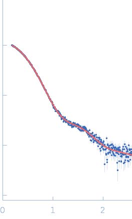
Synchrotron SAXS data from solutions of Lysin from Streptococcus phage P7951 ∆1-208 in 50 mM HEPES, 500 mM NaCl, and 1% glycerol, pH 7 were collected on the BM29 beam line at the ESRF (Grenoble, France) using a Pilatus 1M detector at a sample-detector distance of 2.8 m and at a wavelength of λ = 0.099 nm (I(s) vs s, where s = 4πsinθ/λ, and 2θ is the scattering angle). One solute concentration of 2.70 mg/ml was measured at 20°C. 10 successive 1 second frames were collected. The data were normalized to the intensity of the transmitted beam and radially averaged; the scattering of the solvent-blank was subtracted.
Lysin from Streptococcus phage P7951 (amino acids 209-310) with a C-terminal His-tag.
The bead model displayed in this entry is the volume and bead occupancy-corrected spatial representation of the protein obtained from the spatial alignment of several individual models and does not reflect the fit to the SAXS data.
|
|
![lysin [Streptococcus phage P7951] small angle scattering data](/media/intensities_files/scattering_plots/SASDNU5_dat_img.png) s, nm-1
s, nm-1
![lysin [Streptococcus phage P7951] DAMFILT model](/media/pdb_file/images/SASDNU5_fit1_model1_img.png "Load 3D view")
![lysin [Streptococcus phage P7951] Guinier plot](/media//intensities_files/scattering_plots/SASDNU5_guinier_img.png)
![lysin [Streptococcus phage P7951] Kratky plot](/media/intensities_files/scattering_plots/SASDNU5_kratky_img.png)
![lysin [Streptococcus phage P7951] pair distance distribution function](/media/p_of_R_files/pofr_images/SASDNU5_pofr_img.png)