| MWexperimental | 6 | kDa |
| MWexpected | 6 | kDa |
| VPorod | 8 | nm3 |
|
log I(s)
4.65×10⁻³
4.65×10⁻⁴
4.65×10⁻⁵
4.65×10⁻⁶
|
 s, nm-1
|
|
|
|

|
|

|
|

|
|
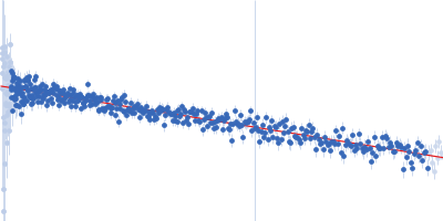
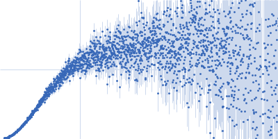
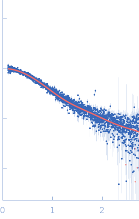
Synchrotron SAXS data from solutions of Monkeypox viral synthetic DNA fragment (MP2 mutant) in 20 mM HEPES, 100 mM KCl, and 0.2 mM EDTA, pH 7.4 were collected on the B21 beam line at the Diamond Light Source (Didcot, UK) using a Eiger 4M detector at a sample-detector distance of 3.7 m and at a wavelength of λ = 0.094 nm (I(s) vs s, where s = 4πsinθ/λ, and 2θ is the scattering angle). In-line size-exclusion chromatography (SEC) SAS was employed. The SEC parameters were as follows: A 40.00 μl sample at 1.0 mg/ml was injected onto a column at 15°C. 600 successive 3 second frames were collected. The data were normalized to the intensity of the transmitted beam and radially averaged; the scattering of the solvent-blank was subtracted.
SEC-column: UNKNOWN: SEC flow-rate: UNKNOWN. |
|
|||||||||||||||||||||