A class III ligand oscillates between internal and terminal binding modes as it engages with the Dishevelled PDZ domain.

Kumar J, Micka M, Komárek J, Klumpler T Bystrý V, Sprangers R, Bařinka C, Bryja V, Tripsianes K, Structure (2025) Europe PMC

SASDWG9 – PDZ domain of the segment polarity protein dishevelled homolog DVL-3

Segment polarity protein dishevelled homolog DVL-3
MWexperimental 10 kDa
MWexpected 12 kDa
VPorod 20 nm3
log I(s) 7.95×10⁻¹ 7.95×10⁻² 7.95×10⁻³ 7.95×10⁻⁴
Segment polarity protein dishevelled homolog DVL-3 small angle scattering data  s, nm-1
ln I(s)
Segment polarity protein dishevelled homolog DVL-3 Guinier plot ln 7.95×10⁻¹ Rg: 1.6 nm 0 (1.6 nm)-2 s2
(sRg)2I(s)/I(0)
Segment polarity protein dishevelled homolog DVL-3 Kratky plot 1.104 0 3 sRg
p(r)
Segment polarity protein dishevelled homolog DVL-3 pair distance distribution function Rg: 1.6 nm 0 Dmax: 4.3 nm

Data validation


Fits and models


log I(s)
 s, nm-1
Segment polarity protein dishevelled homolog DVL-3 REFMAC model

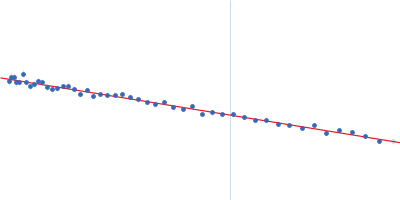
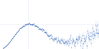
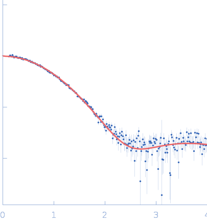
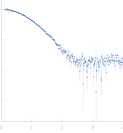
SAXS data from solutions of PDZ domain of the segment polarity protein dishevelled homolog DVL-3 in 50 mM sodium phosphate, 50 mM NaCl, 0.5 mM EDTA, pH 6.5 were collected using a Rigaku BioSAXS-2000 instrument at CEITEC (Brno, Czech Republic) equipped with a Rigaku HyPix-3000 detector at a sample-detector distance of 0.5 m and at a wavelength of λ = 0.154 nm (I(s) vs s, where s = 4πsinθ/λ, and 2θ is the scattering angle). One solute concentration of 5.00 mg/ml was measured at 25°C. Six successive 600 second frames were collected. The data were normalized to the intensity of the transmitted beam and radially averaged; the scattering of the solvent-blank was subtracted.

Segment polarity protein dishevelled homolog DVL-3 (DLV3_PDZ)
Mol. type   Protein
Organism   Homo sapiens
Olig. state   Monomer
Mon. MW   12.0 kDa
 
UniProt   Q92997 (245-351)
Sequence   FASTA