|
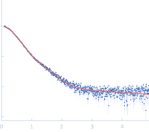
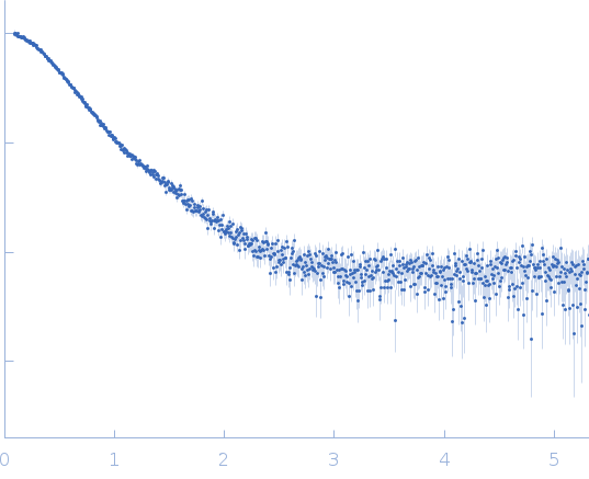
SAXS
data from solutions of
Lipase chaperone (LipH) VD
in
50 mM Tris, 100 mM NaCl, 100 µM TCEP, 5% glycerol,, pH 8
were collected
on the
Xenocs Xeuss 2.0 Q-Xoom instrument (Center for Structural Studies, Heinrich-Heine-University, Düsseldorf, Germany)
using a Pilatus3 R 300K detector
at a sample-detector distance of 0.6 m and
at a wavelength of λ = 0.154 nm
(I(s) vs s, where s = 4πsinθ/λ, and 2θ is the scattering angle).
One solute concentration of 11.82 mg/ml was measured
at 10°C.
24 successive
600 second frames were collected.
The data were normalized to the intensity of the transmitted beam and radially averaged; the scattering of the solvent-blank was subtracted.
|
|
Lipase chaperone
(LipH VD)
|
| Mol. type |
|
Protein |
| Organism |
|
Pseudomonas aeruginosa (strain ATCC 15692 / DSM 22644 / CIP 104116 / JCM 14847 / LMG 12228 / 1C / PRS 101 / PAO1) |
| Olig. state |
|
Monomer |
| Mon. MW |
|
38.3 kDa |
| |
| UniProt |
|
Q01725
|
| Sequence |
|
FASTA |
| |
|
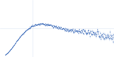
 s, nm-1
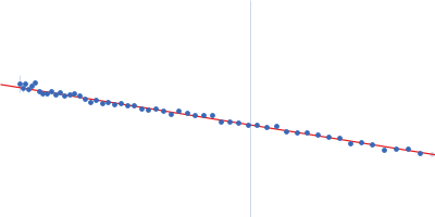
 VD Rg histogram") Rg, nm
Rg, nm