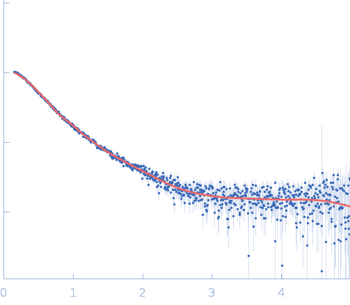
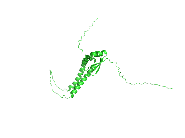
| MWexperimental | 21 | kDa |
| MWexpected | 21 | kDa |
| VPorod | 36 | nm3 |
|
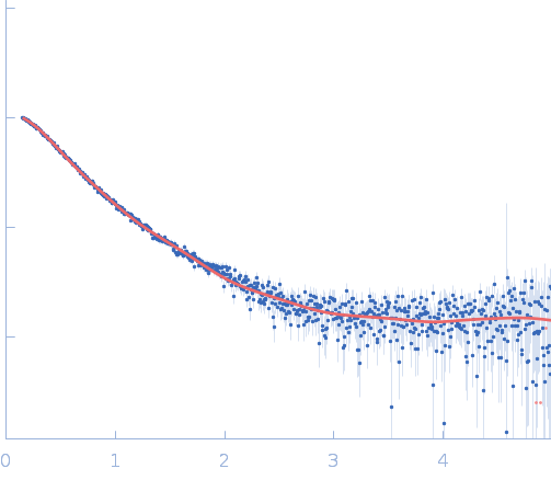
log I(s)
7.87×100
7.87×10⁻¹
7.87×10⁻²
7.87×10⁻³
|
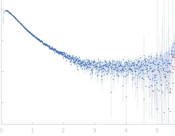 s, nm-1
|
|
|
|
|
|
|
|
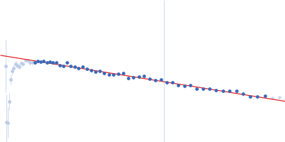
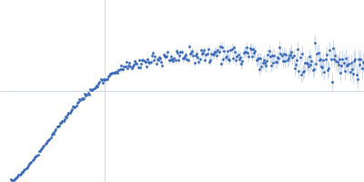
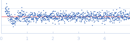
Synchrotron SAXS
data from solutions of
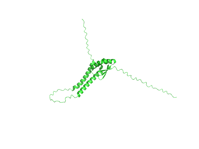
DNA replication protein DciA from M.turberculosis
in
20 mM HEPES, 300 mM NaCl, 2mM b-Me, 5% glycerol, pH 7.5
were collected
on the
BM29 beam line
at the ESRF storage ring
(Grenoble, France)
using a Pilatus3 2M detector
at a sample-detector distance of 2.8 m and
at a wavelength of λ = 0.099 nm
(I(s) vs s, where s = 4πsinθ/λ, and 2θ is the scattering angle).
In-line size-exclusion chromatography (SEC) SAS was employed. The SEC parameters were as follows: A 45.00 μl sample
at 4.3 mg/ml was injected at a 0.50 ml/min flow rate
onto a Agilent AdvanceBio SEC 300Å, 4.6 x 150 mm column
at 20°C.
1600 successive
2 second frames were collected.
The data were normalized to the intensity of the transmitted beam and radially averaged; the scattering of the solvent-blank was subtracted.
|
|
|||||||||||||||||||||