|
Synchrotron SAXS
data from solutions of
hexamer of human citrate synthase and human malate dehydrogenase 2
in
50 mM Tris, 100 mM NaCl, 0.1 mM EDTA, pH 7.5
were collected
on the
12.3.1 (SIBYLS) beam line
at the Advanced Light Source (ALS) storage ring
(Berkeley, CA, USA)
using a Pilatus3 X 2M detector
(I(s) vs s, where s = 4πsinθ/λ, and 2θ is the scattering angle).
In-line size-exclusion chromatography (SEC) SAS was employed. The SEC parameters were as follows: A sample
was injected
onto a column
.
The data were normalized to the intensity of the transmitted beam and radially averaged; the scattering of the solvent-blank was subtracted.
200 μL of 20 mM MES pH 6.4, 900 μL 1.3 mg mL-1 hCS, 300 μL 8.1 mg mL-1 hMDH2, and 1100 μL nanopure H2O were combined and incubated at room temperature for 15 minutes. 300 μL of 0.5% glutaraldehyde stock was combined with the protein solution and rapidly vortexed. The solution was incubated at room temperature for 30 minutes. We added 300 μL of 1 M TRIS base pH 8 to quench excess glutaraldehyde and then incubated the sample at room temperature for approximately 5 minutes. We then purified the sample with an AKTA Start FPLC and a Sephacryl 16/60 S200 size exclusion column in 50 mM Tris-Cl, pH 7.5, 100 mM NaCl, and 0.1 mM EDTA. Fractions containing crosslinked protein were identified with SDS-PAGE and concentrated in 15 mL concentrator tubes at 4ºC.
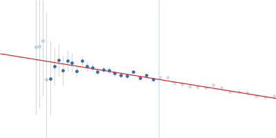
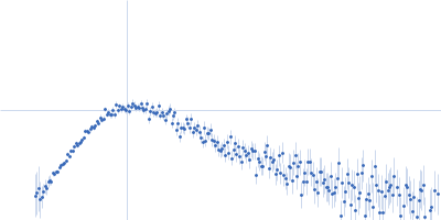
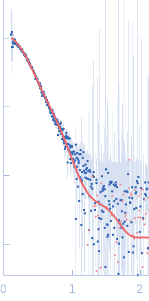
For hCS, we used SEC-SAXS and HT-SAXS to describe the structure, while we used SEC-SAXS for the hMDH2-hCS crosslinked sample. hCS was diluted to a concentration ranging from 0.07 mg mL-1 to 2.0 mg mL-1 in size-exclusion buffer (50 mM Tris-Cl, 100 mM NaCl, and 0.1 mM EDTA, pH 7.5) and then placed into a 96-well plate. The samples were frozen at -80 ºC before shipment. Data were collected at SIBYLS beamline 12.3.1 at the Advanced Light Source, Lawrence Berkeley National Laboratory (Classen et al., 2013; Rosenberg et al., 2022). Before sample collection, the sample plate was spun at 3700 rev min-1 for 10 minutes. Samples were held at 10 ºC during collection. The exposure was 15 seconds, with frames collected every 0.3 seconds for 40 frames per sample. The incident light wavelength was 1.03 Å at a sample-to-detector distance of 2.1 m. This setup results in scattering vectors, q, ranging from 0.013 to 0.5 Å−1, where the scattering vector is defined as q = 4πsinθ/λ, and 2θ is the measured scattering angle.
For SEC-SAXS, samples were separated on a Shodex KW-803 column at a flow rate of 0.5 mL/min at 10 °C, and eluate was measured in-line with UV/vis absorbance at 280 nm, Multi-Angle X-ray Scattering (MALS), and SAXS. The column buffer was 50 mM sodium phosphate, pH 7, 100 mM NaCl, 2 mM DTT, and 2% glycerol. The incident light wavelength was 1.03 Å at a sample-to-detector distance of 2.1 m.
To process SEC-SAXS data, Chromixs was used to subtract buffer from the peaks and produce the initial data file (Panjkovich & Svergun, 2018; Manalastas-Cantos et al., 2021). We then used RAW to analyze the data and generate statistics for the processed SEC-SAXS and HT-SAXS data and FOXS to fit structural models to SAXS data (Schneidman-Duhovny et al., 2013; Hopkins, 2024).
|
|
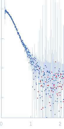 s, nm-1