A class III ligand oscillates between internal and terminal binding modes as it engages with the Dishevelled PDZ domain.

Kumar J, Micka M, Komárek J, Klumpler T, Bystrý V, Sprangers R, Bařinka C, Bryja V, Tripsianes K, Structure (2025) Europe PMC

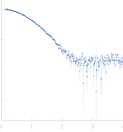
SASDWG9 – PDZ domain of the segment polarity protein dishevelled homolog DVL-3

Segment polarity protein dishevelled homolog DVL-3
MWexperimental 10 kDa
MWexpected 12 kDa
VPorod 20 nm3
log I(s) 7.95×10⁻¹ 7.95×10⁻² 7.95×10⁻³ 7.95×10⁻⁴
Segment polarity protein dishevelled homolog DVL-3 small angle scattering data  s, nm-1
ln I(s)
Segment polarity protein dishevelled homolog DVL-3 Guinier plot ln 7.95×10⁻¹ Rg: 1.6 nm 0 (1.6 nm)-2 s2
(sRg)2I(s)/I(0)
Segment polarity protein dishevelled homolog DVL-3 Kratky plot 1.104 0 3 sRg
p(r)
Segment polarity protein dishevelled homolog DVL-3 pair distance distribution function Rg: 1.6 nm 0 Dmax: 4.3 nm

Data validation


Fits and models


log I(s)
 s, nm-1
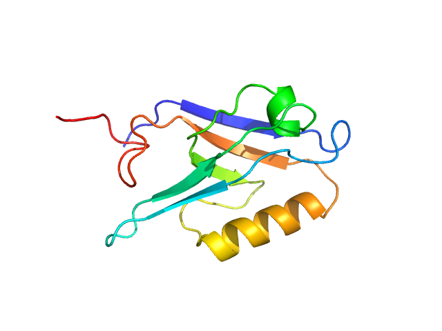
Segment polarity protein dishevelled homolog DVL-3 REFMAC model

SAXS data from solutions of PDZ domain of the segment polarity protein dishevelled homolog DVL-3 in 50 mM sodium phosphate, 50 mM NaCl, 0.5 mM EDTA, pH 6.5 were collected using a Rigaku BioSAXS-2000 instrument at CEITEC (Brno, Czech Republic) equipped with a Rigaku HyPix-3000 detector at a sample-detector distance of 0.5 m and at a wavelength of λ = 0.154 nm (I(s) vs s, where s = 4πsinθ/λ, and 2θ is the scattering angle). One solute concentration of 5.00 mg/ml was measured at 25°C. Six successive 600 second frames were collected. The data were normalized to the intensity of the transmitted beam and radially averaged; the scattering of the solvent-blank was subtracted.

Segment polarity protein dishevelled homolog DVL-3 (DLV3_PDZ)
Mol. type   Protein
Organism   Homo sapiens
Olig. state   Monomer
Mon. MW   12.0 kDa
 
UniProt   Q92997 (245-351)
Sequence   FASTA