The structural basis of fungal glucuronoyl esterase activity on natural substrates.

Ernst HA, Mosbech C, Langkilde AE, Westh P, Meyer AS, Agger JW, Larsen S, Nat Commun 11(1):1026 (2020) Europe PMC

SASDGD6 – Glucuronoyl esterase S286A, deglycolsylated truncated (amino acids 95-474)

4-O-methyl-glucuronoyl methylesterase (Glucuronoyl esterase, truncated)
MWexperimental 35 kDa
MWexpected 43 kDa
VPorod 50 nm3
log I(s) 3.70×10⁻² 3.70×10⁻³ 3.70×10⁻⁴ 3.70×10⁻⁵
4-O-methyl-glucuronoyl methylesterase (Glucuronoyl esterase, truncated) small angle scattering data  s, nm-1
ln I(s)
4-O-methyl-glucuronoyl methylesterase (Glucuronoyl esterase, truncated) Guinier plot ln 3.70×10⁻² Rg: 2.0 nm 0 (2.0 nm)-2 s2
(sRg)2I(s)/I(0)
4-O-methyl-glucuronoyl methylesterase (Glucuronoyl esterase, truncated) Kratky plot 1.104 0 3 sRg
p(r)
4-O-methyl-glucuronoyl methylesterase (Glucuronoyl esterase, truncated) pair distance distribution function Rg: 2.1 nm 0 Dmax: 6.1 nm

Data validation


Fits and models


log I(s)
 s, nm-1
4-O-methyl-glucuronoyl methylesterase (Glucuronoyl esterase, truncated) PDB (PROTEIN DATA BANK) model

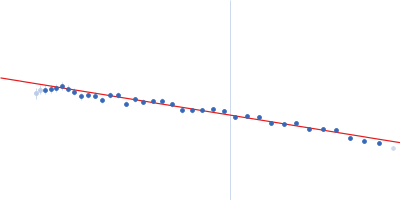
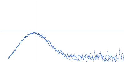
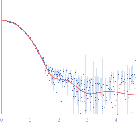
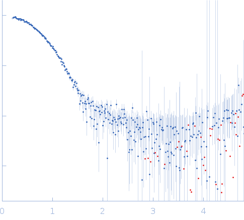
SAXS data from solutions of N-terminal truncated mutant glucuronoyl esterase (S286A, deglycolsylated) in 20 mM sodium acetate, pH 5 were collected using a Xenocs BioXolver L with GeniX3D (University of Copenhagen, Department of Drug Design and Pharmacology) equipped with a Pilatus3 R 300K detector at a sample-detector distance of 0.6 m and at a wavelength of λ = 0.154 nm (I(s) vs s, where s = 4πsinθ/λ, and 2θ is the scattering angle). One solute concentration of 3.60 mg/ml was measured at 21°C. 10 successive 60 second frames were collected. The data were normalized to the intensity of the transmitted beam and radially averaged; the scattering of the solvent-blank was subtracted.

4-O-methyl-glucuronoyl methylesterase (Glucuronoyl esterase, truncated) (CuGE)
Mol. type   Protein
Organism   Cerrena unicolor
Olig. state   Monomer
Mon. MW   43.2 kDa
 
UniProt   A0A0A7EQR3 (95-474)
Sequence   FASTA
 
PDB ID   6RTV