| MWexperimental | 98 | kDa |
| MWexpected | 100 | kDa |
|
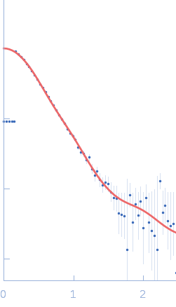
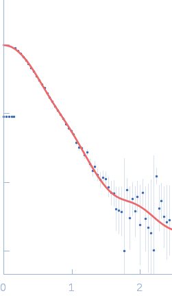
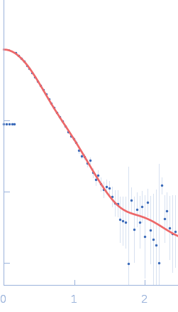
log I(s)
4.10×100
4.10×10⁻¹
4.10×10⁻²
4.10×10⁻³
|
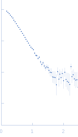 s, nm-1
|
|
|
|

|
|

|
|

|
|
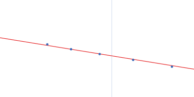
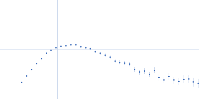
Synchrotron SAXS data from solutions of the multi-component estrogen receptor in 10 mM CHES, 125 mM NaCl, 5mM KCl, 4 mM MgCl2, 50 mM arginine, 50 mM glutamate, 5 mM TCEP, 5% glycerol, 10 µm Zn acetate, 10 µM estradiol, pH 9.5 were collected on the BioCAT 18ID beam line at the Advanced Photon Source (APS; Argonne, IL, USA) using a MAR 165 CCD detector at a sample-detector distance of 3 m and at a wavelength of λ = 0.103 nm (l(s) vs s, where s = 4πsinθ/λ, and 2θ is the scattering angle). Solute concentrations ranging between 0.1 and 0.8 mg/ml were measured at 10°C from size-exclusion chromatography eluates. 20 successive 1.100 second frames were collected. The data were normalized to the intensity of the transmitted beam and radially averaged; the scattering of the solvent-blank was subtracted.
Additional SEC parameters: Column type: GE Healthcare Superdex 200 GL 10/300; Flow rate: 0.5 ml/min; Sample injection concentration: 3.5 mg/ml; Injection volume: 500 µl. |
|
||||||||||||||||||||||||||||||||||||||||||||||||||||||||||||||||||||||||||||||||||||||||||||||||||||||||||||||||||||||||