|
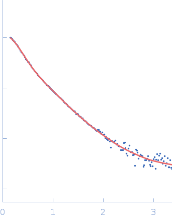
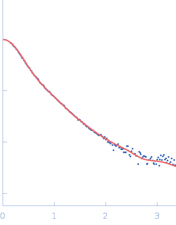
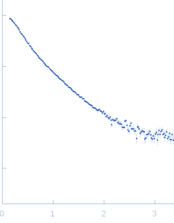
Synchrotron SAXS data from solutions of the p67phox subunit of phagocyte NADPH oxidase in 20 mM HEPES, 50 mM NaCl, 1 mM EDTA, 2 mM DTT, 5% glycerol, pH 8 were collected on the D24 beam line at the LURE storage ring (Orsay, France) using a linear gas home-made detector at a sample-detector distance of 1.4 m and at a wavelength of λ = 0.1488 nm (l(s) vs s, where s = 4πsinθ/λ, and 2θ is the scattering angle). Samples were studied at different concentrations ranging from 0.3 to 3.8 mg/ml measured at 4°C. Eight successive 200 second frames were collected. SAXS data were normalized to the intensity of the incident beam, averaged and background subtracted using the program package PRIMUS. Intensities were put on an absolute scale (cm-1) using water scattering. The final pattern used for fitting was obtained by extrapolating to infinite dilution of the set of curves recorded at the different concentrations. Molecular mass was obtained using the bayesian inference approach proposed in PRIMUS/qt, which combines four concentration independent MM estimators: M=62.4 kDa within the credibility interval [60.2-67.9].
|
|
 s, nm-1
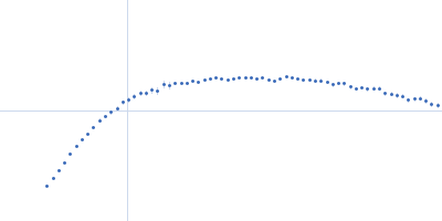
 subunit of phagocyte NADPH oxidase Rg histogram") Rg, nm
Rg, nm