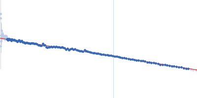
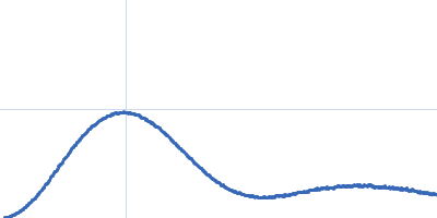
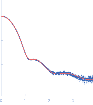
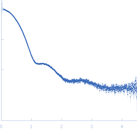
Structural characterization of a Vatairea macrocarpa lectin in complex with a tumor-associated antigen: A new tool for cancer research.

Sousa BL, Silva-Filho JC, Kumar P, Graewert MA, Pereira RI, Cunha RMS, Nascimento KS, Bezerra GA, Delatorre P, Djinovic-Carugo K, Nagano CS, Gruber K, Cavada BS, Int J Biochem Cell Biol 72:27-39 (2016) Europe PMC

SASDBS2 – Recombinant Tn antigen-binding lectin from Vatairea macrocarpa

Recombinant Tn antigen-binding lectin
MWI(0) 70 kDa
MWexpected 105 kDa
VPorod 168 nm3
log I(s) 1.81×10³ 1.81×10² 1.81×101 1.81×100
Recombinant Tn antigen-binding lectin small angle scattering data  s, nm-1
ln I(s)
Recombinant Tn antigen-binding lectin Guinier plot ln 1.81×10³ Rg: 3.2 nm 0 (3.2 nm)-2 s2
(sRg)2I(s)/I(0)
Recombinant Tn antigen-binding lectin Kratky plot 1.104 0 3 sRg
p(r)
Recombinant Tn antigen-binding lectin pair distance distribution function Rg: 3.1 nm 0 Dmax: 9.5 nm

Data validation


Fits and models


log I(s)
 s, nm-1
Recombinant Tn antigen-binding lectin CORAL model

Synchrotron SAXS data from solutions of Recombinant Tn antigen-binding lectin from Vatairea macrocarpa in 100 mM sodium phosphate 150 mM NaCl 5% (v/v) glycerol, pH 5 were collected on the EMBL P12 beam line at the PETRA III storage ring (DESY; Hamburg, Germany) using a Pilatus 2M detector at a sample-detector distance of 3.1 m and at a wavelength of λ = 0.12 nm (I(s) vs s, where s = 4πsinθ/λ, and 2θ is the scattering angle). Solute concentrations ranging between 1 and 10 mg/ml were measured . 20 successive 0.050 second frames were collected. The data were normalized to the intensity of the transmitted beam and radially averaged; the scattering of the solvent-blank was subtracted.

Note: Carries a tyrosine mutation, instead of phenylalanine, at position 6 (F6Y - cf UniProt P81371).

Recombinant Tn antigen-binding lectin (vRML)
Mol. type   Protein
Organism   Vatairea macrocarpa
Olig. state   Tetramer
Mon. MW   26.2 kDa
 
UniProt   P81371
Sequence   FASTA