|
|
|
|
|
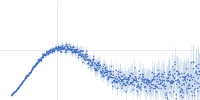
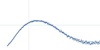
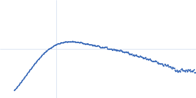
| Sample: |
Endo-beta-N-acetylglucosaminidase H dimer, 61 kDa Streptomyces plicatus protein
|
| Buffer: |
20 mM Tris-HCl, 50 mM NaCl, 5 mM EDTA, pH: 7.5 |
| Experiment: |
SAXS
data collected at 12.3.1 (SIBYLS), Advanced Light Source (ALS) on 2019 Nov 4
|
SAXS studies of X-ray induced disulfide bond damage: Engineering high-resolution insight from a low-resolution technique
PLOS ONE 15(11):e0239702 (2020)
Stachowski T, Snell M, Snell E, Boggon T
|
| RgGuinier |
2.8 |
nm |
| VolumePorod |
60 |
nm3 |
|
|
|
|
|
|
|
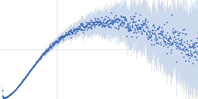
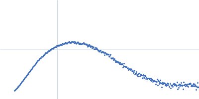
| Sample: |
Endo-beta-N-acetylglucosaminidase H dimer, 61 kDa Streptomyces plicatus protein
|
| Buffer: |
20 mM Tris-HCl, 50 mM NaCl, 5 mM EDTA, pH: 7.5 |
| Experiment: |
SAXS
data collected at 12.3.1 (SIBYLS), Advanced Light Source (ALS) on 2019 Nov 4
|
SAXS studies of X-ray induced disulfide bond damage: Engineering high-resolution insight from a low-resolution technique
PLOS ONE 15(11):e0239702 (2020)
Stachowski T, Snell M, Snell E, Boggon T
|
| RgGuinier |
2.7 |
nm |
| VolumePorod |
60 |
nm3 |
|
|
|
|
|
|
|
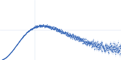
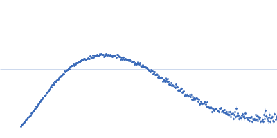
| Sample: |
Endo-beta-N-acetylglucosaminidase H dimer, 61 kDa Streptomyces plicatus protein
|
| Buffer: |
20 mM Tris-HCl, 50 mM NaCl, 5 mM EDTA, pH: 7.5 |
| Experiment: |
SAXS
data collected at 12.3.1 (SIBYLS), Advanced Light Source (ALS) on 2019 Nov 4
|
SAXS studies of X-ray induced disulfide bond damage: Engineering high-resolution insight from a low-resolution technique
PLOS ONE 15(11):e0239702 (2020)
Stachowski T, Snell M, Snell E, Boggon T
|
| RgGuinier |
2.7 |
nm |
| VolumePorod |
61 |
nm3 |
|
|
|
|
|
|
|
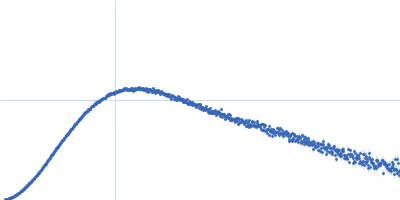
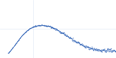
| Sample: |
Endo-beta-N-acetylglucosaminidase H dimer, 61 kDa Streptomyces plicatus protein
|
| Buffer: |
20 mM Tris-HCl, 50 mM NaCl, 5 mM EDTA, pH: 7.5 |
| Experiment: |
SAXS
data collected at 12.3.1 (SIBYLS), Advanced Light Source (ALS) on 2019 Nov 4
|
SAXS studies of X-ray induced disulfide bond damage: Engineering high-resolution insight from a low-resolution technique
PLOS ONE 15(11):e0239702 (2020)
Stachowski T, Snell M, Snell E, Boggon T
|
| RgGuinier |
2.8 |
nm |
| VolumePorod |
61 |
nm3 |
|
|
|
|
|
|
|
| Sample: |
Endo-beta-N-acetylglucosaminidase H dimer, 61 kDa Streptomyces plicatus protein
|
| Buffer: |
20 mM Tris-HCl, 50 mM NaCl, 5 mM EDTA, pH: 7.5 |
| Experiment: |
SAXS
data collected at 12.3.1 (SIBYLS), Advanced Light Source (ALS) on 2019 Nov 4
|
SAXS studies of X-ray induced disulfide bond damage: Engineering high-resolution insight from a low-resolution technique
PLOS ONE 15(11):e0239702 (2020)
Stachowski T, Snell M, Snell E, Boggon T
|
| RgGuinier |
2.7 |
nm |
| VolumePorod |
60 |
nm3 |
|
|
|
|
|
|
|
| Sample: |
Candidatus Glomeribacter gigasporarum cyclodipeptide synthase monomer, 34 kDa Candidatus Glomeribacter gigasporarum protein
E. coli Phe-tRNAPhe monomer, 25 kDa Escherichia coli RNA
|
| Buffer: |
10 mM MOPS pH6.7; 200 mM NaCl, 8 mM MgCl2, pH: 6.7 |
| Experiment: |
SAXS
data collected at SWING, SOLEIL on 2016 Oct 2
|
Structural basis of the interaction between cyclodipeptide synthases and aminoacylated tRNA substrates.
RNA 26(11):1589-1602 (2020)
Bourgeois G, Seguin J, Babin M, Gondry M, Mechulam Y, Schmitt E
|
| RgGuinier |
3.3 |
nm |
| Dmax |
14.0 |
nm |
| VolumePorod |
77 |
nm3 |
|
|
|
|
|
|
|
| Sample: |
ACT domain of Rel protein (Bifunctional (p)ppGpp synthase/hydrolase RelA) dimer, 20 kDa Mycobacterium tuberculosis protein
|
| Buffer: |
50 mM Tris-HCl, 350 mM NaCl, 5% glycerol, 1 mM DTT, pH: 8.5 |
| Experiment: |
SAXS
data collected at Bruker Nanostar, Nanyang Technological University on 2018 Jun 7
|
Atomic structure of, and valine binding to the regulatory ACT domain of the Mycobacterium tuberculosis Rel protein.
FEBS J (2020)
Shin J, Singal B, Manimekalai MSS, Chen MW, Ragunathan P, Grüber G
|
| RgGuinier |
1.9 |
nm |
| Dmax |
6.1 |
nm |
| VolumePorod |
29 |
nm3 |
|
|
|
|
|
|
|
| Sample: |
Myosin essential light chain monomer, 16 kDa Plasmodium falciparum protein
|
| Buffer: |
20 mM HEPES pH 7.5, 150 mM NaCl, 0.5 mM TCEP, pH: 7.5 |
| Experiment: |
SAXS
data collected at EMBL P12, PETRA III on 2018 Jun 30
|
Structural role of essential light chains in the apicomplexan glideosome.
Commun Biol 3(1):568 (2020)
Pazicky S, Dhamotharan K, Kaszuba K, Mertens HDT, Gilberger T, Svergun D, Kosinski J, Weininger U, Löw C
|
| RgGuinier |
2.7 |
nm |
| Dmax |
9.5 |
nm |
| VolumePorod |
23 |
nm3 |
|
|
|
|
|
|
|
| Sample: |
Myosin essential light chain 2 monomer, 15 kDa Toxoplasma gondii protein
|
| Buffer: |
20 mM HEPES pH 7.5, 150 mM NaCl, 0.5 mM TCEP, pH: 7.5 |
| Experiment: |
SAXS
data collected at EMBL P12, PETRA III on 2019 Apr 8
|
Structural role of essential light chains in the apicomplexan glideosome.
Commun Biol 3(1):568 (2020)
Pazicky S, Dhamotharan K, Kaszuba K, Mertens HDT, Gilberger T, Svergun D, Kosinski J, Weininger U, Löw C
|
| RgGuinier |
2.1 |
nm |
| Dmax |
6.7 |
nm |
| VolumePorod |
27 |
nm3 |
|
|
|
|
|
|
|
| Sample: |
Myosin essential light chain monomer, 16 kDa Plasmodium falciparum protein
Plasmodium falciparum myosin A monomer, 5 kDa Plasmodium falciparum protein
Myosin A tail domain interacting protein monomer, 17 kDa Plasmodium falciparum protein
|
| Buffer: |
20 mM HEPES pH 7.5, 150 mM NaCl, 0.5 mM TCEP, pH: 7.5 |
| Experiment: |
SAXS
data collected at EMBL P12, PETRA III on 2019 Sep 30
|
Structural role of essential light chains in the apicomplexan glideosome.
Commun Biol 3(1):568 (2020)
Pazicky S, Dhamotharan K, Kaszuba K, Mertens HDT, Gilberger T, Svergun D, Kosinski J, Weininger U, Löw C
|
| RgGuinier |
2.7 |
nm |
| Dmax |
10.7 |
nm |
| VolumePorod |
51 |
nm3 |
|
|


ppGpp synthase/hydrolase RelA) experimental SAS data")