|
|
|
|
|
| Sample: |
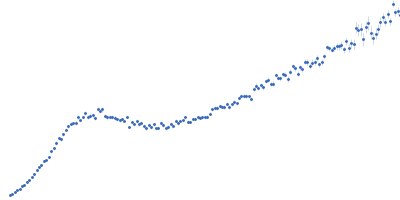
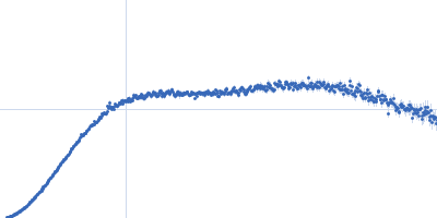
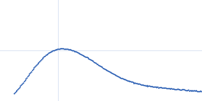
Ferredoxin protease E83A mutant monomer, 101 kDa Pectobacterium atrosepticum SCRI1043 protein
Arabidopsis ferredoxin 2 monomer, 11 kDa Arabidopsis thaliana protein
|
| Buffer: |
20 mM Tris, 150 mM NaCl, pH: 7.8 |
| Experiment: |
SAXS
data collected at SAXS/WAXS, Australian Synchrotron on 2017 Oct 26
|
FusC, a member of the M16 protease family acquired by bacteria for iron piracy against plants.
PLoS Biol 16(8):e2006026 (2018)
Grinter R, Hay ID, Song J, Wang J, Teng D, Dhanesakaran V, Wilksch JJ, Davies MR, Littler D, Beckham SA, Henderson IR, Strugnell RA, Dougan G, Lithgow T
|
| RgGuinier |
3.8 |
nm |
| Dmax |
14.2 |
nm |
| VolumePorod |
178 |
nm3 |
|
|
|
|
|
|
|
| Sample: |
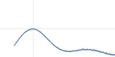
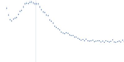
Ferredoxin protease E83A mutant monomer, 101 kDa Pectobacterium atrosepticum SCRI1043 protein
Arabidopsis ferredoxin 2 monomer, 11 kDa Arabidopsis thaliana protein
|
| Buffer: |
20 mM Tris, 150 mM NaCl, pH: 7.8 |
| Experiment: |
SAXS
data collected at SAXS/WAXS, Australian Synchrotron on 2017 Oct 26
|
FusC, a member of the M16 protease family acquired by bacteria for iron piracy against plants.
PLoS Biol 16(8):e2006026 (2018)
Grinter R, Hay ID, Song J, Wang J, Teng D, Dhanesakaran V, Wilksch JJ, Davies MR, Littler D, Beckham SA, Henderson IR, Strugnell RA, Dougan G, Lithgow T
|
| RgGuinier |
3.7 |
nm |
| Dmax |
14.0 |
nm |
| VolumePorod |
175 |
nm3 |
|
|
|
|
|
|
|
| Sample: |
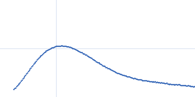
Ferredoxin protease E83A mutant monomer, 101 kDa Pectobacterium atrosepticum SCRI1043 protein
Arabidopsis ferredoxin 2 monomer, 11 kDa Arabidopsis thaliana protein
|
| Buffer: |
20 mM Tris, 150 mM NaCl, pH: 7.8 |
| Experiment: |
SAXS
data collected at SAXS/WAXS, Australian Synchrotron on 2017 Oct 26
|
FusC, a member of the M16 protease family acquired by bacteria for iron piracy against plants.
PLoS Biol 16(8):e2006026 (2018)
Grinter R, Hay ID, Song J, Wang J, Teng D, Dhanesakaran V, Wilksch JJ, Davies MR, Littler D, Beckham SA, Henderson IR, Strugnell RA, Dougan G, Lithgow T
|
| RgGuinier |
3.7 |
nm |
| Dmax |
14.0 |
nm |
| VolumePorod |
180 |
nm3 |
|
|
|
|
|
|
|
| Sample: |
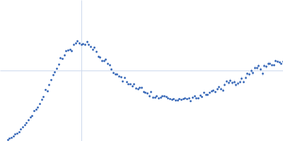
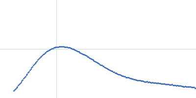
Ovalbumin monomer, 43 kDa Gallus gallus protein
|
| Buffer: |
Water, pH: 7 |
| Experiment: |
SAXS
data collected at Bruker Nonius FR591, University of Pennslyvania on 2013 Jun 27
|
The Proof Is in the Pidan: Generalizing Proteins as Patchy Particles.
ACS Cent Sci 4(7):840-853 (2018)
Cai J, Sweeney AM
|
|
|
|
|
|
|
|
| Sample: |
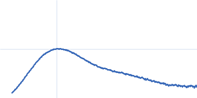
Ovalbumin (common quail) monomer, 42 kDa Coturnix coturnix protein
|
| Buffer: |
Water, pH: 7 |
| Experiment: |
SAXS
data collected at Bruker Nonius FR591, University of Pennslyvania on 2013 Jun 27
|
The Proof Is in the Pidan: Generalizing Proteins as Patchy Particles.
ACS Cent Sci 4(7):840-853 (2018)
Cai J, Sweeney AM
|
|
|
|
|
|
|
|
| Sample: |
Ovalbumin monomer, 43 kDa Gallus gallus protein
|
| Buffer: |
Water, pH: 7 |
| Experiment: |
SAXS
data collected at Bruker Nonius FR591, University of Pennslyvania on 2013 Jun 27
|
The Proof Is in the Pidan: Generalizing Proteins as Patchy Particles.
ACS Cent Sci 4(7):840-853 (2018)
Cai J, Sweeney AM
|
|
|
|
|
|
|
|
| Sample: |
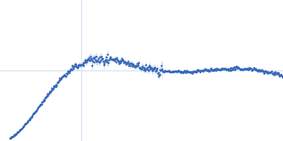
Anti-CD32b Antibody Clone 6G08 Antibody Binding Fragment monomer, 46 kDa Homo sapiens protein
|
| Buffer: |
50mM HEPES, 150mM KCl, pH: 7.5 |
| Experiment: |
SAXS
data collected at BM29, ESRF on 2014 Dec 5
|
Evaluating Anti-CD32b F(ab) Conformation Using Molecular Dynamics and Small-Angle X-Ray Scattering.
Biophys J 115(2):289-299 (2018)
Sutton EJ, Bradshaw RT, Orr CM, Frendéus B, Larsson G, Teige I, Cragg MS, Tews I, Essex JW
|
| RgGuinier |
2.6 |
nm |
| Dmax |
7.3 |
nm |
| VolumePorod |
68 |
nm3 |
|
|
|
|
|
|
|
| Sample: |
Small glutamine-rich tetratricopeptide repeat-containing protein alpha full length dimer, 68 kDa Homo sapiens protein
|
| Buffer: |
10 mM potassium phosphate, 100 mM NaCl, pH: 6 |
| Experiment: |
SAXS
data collected at EMBL P12, PETRA III on 2015 Jun 5
|
Structural complexity of the co-chaperone SGTA: a conserved C-terminal region is implicated in dimerization and substrate quality control.
BMC Biol 16(1):76 (2018)
Martínez-Lumbreras S, Krysztofinska EM, Thapaliya A, Spilotros A, Matak-Vinkovic D, Salvadori E, Roboti P, Nyathi Y, Muench JH, Roessler MM, Svergun DI, High S, Isaacson RL
|
|
|
|
|
|
|
|
| Sample: |
Small glutamine-rich tetratricopeptide repeat-containing protein alpha Nterminal-TPR domains dimer, 47 kDa Homo sapiens protein
|
| Buffer: |
10 mM potassium phosphate, 100 mM NaCl, pH: 6 |
| Experiment: |
SAXS
data collected at EMBL P12, PETRA III on 2015 Jun 5
|
Structural complexity of the co-chaperone SGTA: a conserved C-terminal region is implicated in dimerization and substrate quality control.
BMC Biol 16(1):76 (2018)
Martínez-Lumbreras S, Krysztofinska EM, Thapaliya A, Spilotros A, Matak-Vinkovic D, Salvadori E, Roboti P, Nyathi Y, Muench JH, Roessler MM, Svergun DI, High S, Isaacson RL
|
|
|
|
|
|
|
|
| Sample: |
4-hydroxy-tetrahydrodipicolinate synthase from Clostridium botulinum tetramer, 126 kDa Clostridium botulinum protein
|
| Buffer: |
20mM Tris, 150mM NaCl, pH: 8 |
| Experiment: |
SAXS
data collected at SAXS/WAXS, Australian Synchrotron on 2010 Nov 26
|
Substrate Locking Promotes Dimer-Dimer Docking of an Enzyme Antibiotic Target.
Structure 26(7):948-959.e5 (2018)
Atkinson SC, Dogovski C, Wood K, Griffin MDW, Gorman MA, Hor L, Reboul CF, Buckle AM, Wuttke J, Parker MW, Dobson RCJ, Perugini MA
|
| RgGuinier |
3.2 |
nm |
| Dmax |
9.0 |
nm |
| VolumePorod |
159 |
nm3 |
|
|


 experimental SAS data")