|
|
|
|
|
| Sample: |
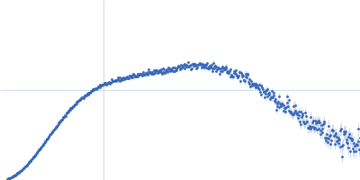
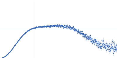
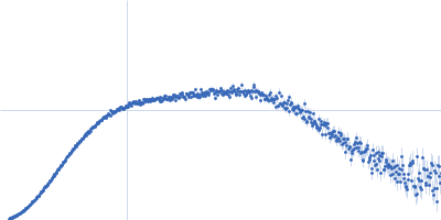
Human telomeric g-quadruplex, 16 kDa DNA
|
| Buffer: |
5 mM KH2PO4/K2HPO4, pH: 7 |
| Experiment: |
SAXS
data collected at BM29, ESRF on 2024 Jul 12
|
Ligands reshape the compactness, stability, and topology of telomeric G-quadruplex dimers.
Nucleic Acids Res 54(8) (2026)
Bertini L, Libera V, Arciuolo V, Trapella M, Marzano S, Mostarac D, Schirò G, Petrillo C, Giancola C, De Michele C, Amato J, Comez L, Pagano B, Paciaroni A
|
|
|
|
|
|
|
|
| Sample: |
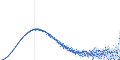
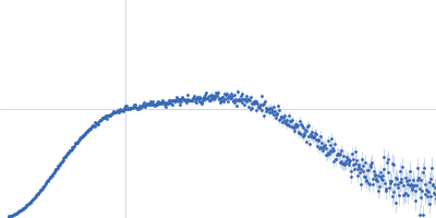
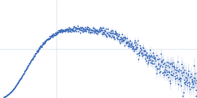
Human telomeric g-quadruplex monomer, 8 kDa DNA
|
| Buffer: |
5 mM KH2PO4/K2HPO4, pH: 7 |
| Experiment: |
SAXS
data collected at BM29, ESRF on 2024 Jul 12
|
Ligands reshape the compactness, stability, and topology of telomeric G-quadruplex dimers.
Nucleic Acids Res 54(8) (2026)
Bertini L, Libera V, Arciuolo V, Trapella M, Marzano S, Mostarac D, Schirò G, Petrillo C, Giancola C, De Michele C, Amato J, Comez L, Pagano B, Paciaroni A
|
|
|
|
|
|
|
|
| Sample: |
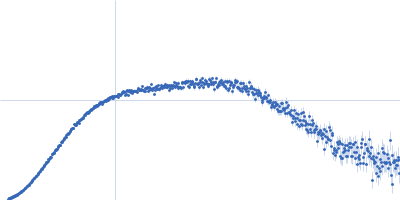
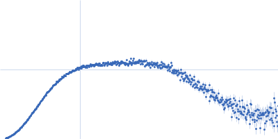
Human telomeric g-quadruplex, 16 kDa DNA
|
| Buffer: |
5 mM KH2PO4/K2HPO4, pH: 7 |
| Experiment: |
SAXS
data collected at BM29, ESRF on 2024 Jul 12
|
Ligands reshape the compactness, stability, and topology of telomeric G-quadruplex dimers.
Nucleic Acids Res 54(8) (2026)
Bertini L, Libera V, Arciuolo V, Trapella M, Marzano S, Mostarac D, Schirò G, Petrillo C, Giancola C, De Michele C, Amato J, Comez L, Pagano B, Paciaroni A
|
|
|
|
|
|
|
|
| Sample: |
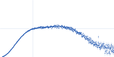
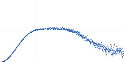
Human telomeric g-quadruplex, 16 kDa DNA
|
| Buffer: |
5 mM KH2PO4/K2HPO4, pH: 7 |
| Experiment: |
SAXS
data collected at BM29, ESRF on 2024 Jul 12
|
Ligands reshape the compactness, stability, and topology of telomeric G-quadruplex dimers.
Nucleic Acids Res 54(8) (2026)
Bertini L, Libera V, Arciuolo V, Trapella M, Marzano S, Mostarac D, Schirò G, Petrillo C, Giancola C, De Michele C, Amato J, Comez L, Pagano B, Paciaroni A
|
|
|