|
|
|
|
|
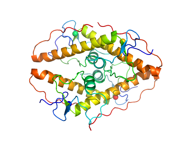
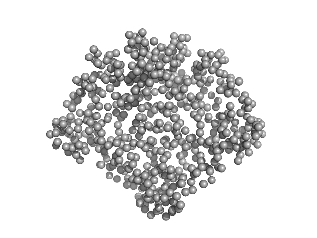
| Sample: |
LD15650p (Pita, isoform A) tetramer, 52 kDa Drosophila melanogaster protein
|
| Buffer: |
20 mM Tris, 100 mM NaCl, 5 mM DTT, pH: 7.4
|
| Experiment: |
SAXS
data collected at BM29, ESRF on 2018 Jul 8
|
Structural insights into highly similar spatial organization of zinc-finger associated domains with a very low sequence similarity.
Structure (2022)
Bonchuk AN, Boyko KM, Nikolaeva AY, Burtseva AD, Popov VO, Georgiev PG
|
| RgGuinier |
2.4 |
nm |
| Dmax |
7.6 |
nm |
| VolumePorod |
62 |
nm3 |
|
|
|
|
|
|
|
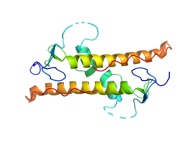
| Sample: |
LD15650p (Pita, isoform A; L45A) dimer, 26 kDa Drosophila melanogaster protein
|
| Buffer: |
20 mM Tris, 100 mM NaCl, 5 mM DTT, pH: 7.4
|
| Experiment: |
SAXS
data collected at BM29, ESRF on 2018 Dec 17
|
Structural insights into highly similar spatial organization of zinc-finger associated domains with a very low sequence similarity.
Structure (2022)
Bonchuk AN, Boyko KM, Nikolaeva AY, Burtseva AD, Popov VO, Georgiev PG
|
| RgGuinier |
2.3 |
nm |
| Dmax |
9.5 |
nm |
| VolumePorod |
35 |
nm3 |
|
|
|
|
|
|
|
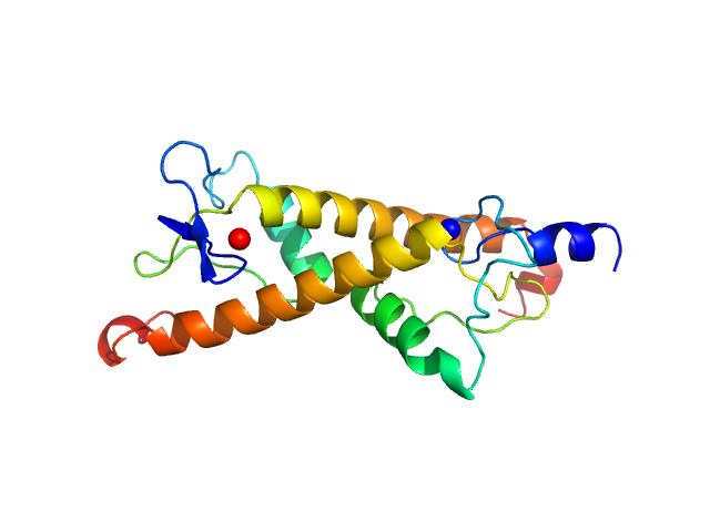
| Sample: |
LD30467p (Motif 1 binding protein) dimer, 22 kDa Drosophila melanogaster protein
|
| Buffer: |
20 mM Bis-Tris-Propane, 100 mM NaCl, 5 mM DTT, pH: 8.5
|
| Experiment: |
SAXS
data collected at BM29, ESRF on 2018 Dec 17
|
Structural insights into highly similar spatial organization of zinc-finger associated domains with a very low sequence similarity.
Structure (2022)
Bonchuk AN, Boyko KM, Nikolaeva AY, Burtseva AD, Popov VO, Georgiev PG
|
| RgGuinier |
2.3 |
nm |
| Dmax |
9.1 |
nm |
| VolumePorod |
33 |
nm3 |
|
|
|
|
|
|
|
| Sample: |
Ganglioside-induced differentiation-associated protein 1 (H123R) dimer, 70 kDa Homo sapiens protein
|
| Buffer: |
25 mM HEPES, 300 mM NaCl, pH: 7.5
|
| Experiment: |
SAXS
data collected at SWING, SOLEIL on 2020 Oct 8
|
Structural insights into Charcot-Marie-Tooth disease-linked mutations in human GDAP1.
FEBS Open Bio (2022)
Sutinen A, Nguyen GTT, Raasakka A, Muruganandam G, Loris R, Ylikallio E, Tyynismaa H, Bartesaghi L, Ruskamo S, Kursula P
|
| RgGuinier |
3.1 |
nm |
| Dmax |
9.9 |
nm |
| VolumePorod |
107 |
nm3 |
|
|
|
|
|
|
|
| Sample: |
Ganglioside-induced differentiation-associated protein 1 (R120W) dimer, 70 kDa Homo sapiens protein
|
| Buffer: |
25 mM HEPES, 300 mM NaCl, pH: 7.5
|
| Experiment: |
SAXS
data collected at SWING, SOLEIL on 2020 Jul 1
|
Structural insights into Charcot-Marie-Tooth disease-linked mutations in human GDAP1.
FEBS Open Bio (2022)
Sutinen A, Nguyen GTT, Raasakka A, Muruganandam G, Loris R, Ylikallio E, Tyynismaa H, Bartesaghi L, Ruskamo S, Kursula P
|
| RgGuinier |
3.1 |
nm |
| Dmax |
10.0 |
nm |
| VolumePorod |
105 |
nm3 |
|
|
|
|
|
|
|
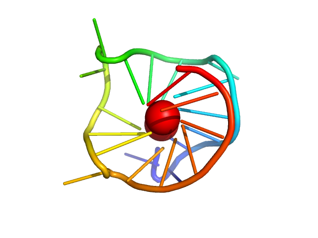
| Sample: |
MYC22-G14T/G23T monomer, 7 kDa Homo sapiens DNA
|
| Buffer: |
50 mM Tris, 30 mM KCl, pH: 8
|
| Experiment: |
SAXS
data collected at BL-10C, Photon Factory (PF), High Energy Accelerator Research Organization (KEK) on 2022 Feb 27
|
Small-angle X-ray scattering data of a guanine-rich DNA derived from the promoter region of c-MYC gene in solution
Data in Brief :108285 (2022)
Miyauchi K, Imamura H, Yamaoki Y, Kato M
|
| RgGuinier |
1.3 |
nm |
| Dmax |
5.3 |
nm |
| VolumePorod |
9 |
nm3 |
|
|
|
|
|
|
|
| Sample: |
MYC22-G14T/G23T monomer, 7 kDa Homo sapiens DNA
|
| Buffer: |
50 mM Tris, 100 mM 18-crown-6, pH: 8
|
| Experiment: |
SAXS
data collected at BL-10C, Photon Factory (PF), High Energy Accelerator Research Organization (KEK) on 2022 Feb 27
|
Small-angle X-ray scattering data of a guanine-rich DNA derived from the promoter region of c-MYC gene in solution
Data in Brief :108285 (2022)
Miyauchi K, Imamura H, Yamaoki Y, Kato M
|
| RgGuinier |
1.6 |
nm |
| Dmax |
6.0 |
nm |
| VolumePorod |
10 |
nm3 |
|
|
|
|
|
|
|
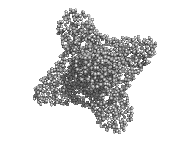
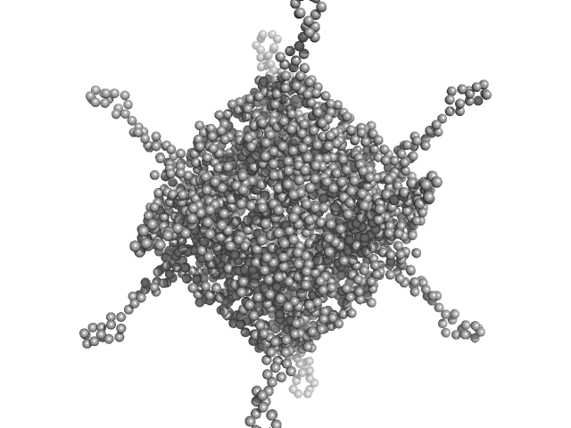
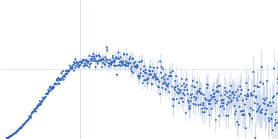
| Sample: |
DNA protection during starvation, DPS (Ferritin superfamily) dodecamer, 270 kDa Deinococcus grandis protein
|
| Buffer: |
50 mM MOPS-NaOH, 50 mM NaCl, pH: 7
|
| Experiment: |
SAXS
data collected at EMBL P12, PETRA III on 2020 Oct 22
|
The Conformation of the N-Terminal Tails of Deinococcus grandis Dps Is Modulated by the Ionic Strength
International Journal of Molecular Sciences 23(9):4871 (2022)
Guerra J, Blanchet C, Vieira B, Almeida A, Waerenborgh J, Jones N, Hoffmann S, Tavares P, Pereira A
|
| RgGuinier |
4.4 |
nm |
| Dmax |
16.1 |
nm |
| VolumePorod |
409 |
nm3 |
|
|
|
|
|
|
|
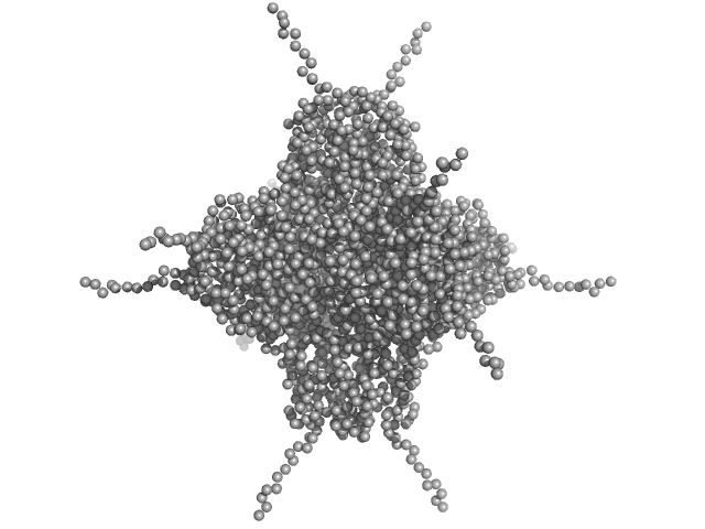
| Sample: |
DNA protection during starvation, DPS (Ferritin superfamily) dodecamer, 270 kDa Deinococcus grandis protein
|
| Buffer: |
50 mM MOPS-NaOH, 80 mM NaCl, pH: 7
|
| Experiment: |
SAXS
data collected at EMBL P12, PETRA III on 2020 Oct 22
|
The Conformation of the N-Terminal Tails of Deinococcus grandis Dps Is Modulated by the Ionic Strength
International Journal of Molecular Sciences 23(9):4871 (2022)
Guerra J, Blanchet C, Vieira B, Almeida A, Waerenborgh J, Jones N, Hoffmann S, Tavares P, Pereira A
|
| RgGuinier |
4.5 |
nm |
| Dmax |
17.1 |
nm |
| VolumePorod |
475 |
nm3 |
|
|
|
|
|
|
|
| Sample: |
DNA protection during starvation, DPS (Ferritin superfamily) dodecamer, 270 kDa Deinococcus grandis protein
|
| Buffer: |
50 mM MOPS-NaOH, 230 mM NaCl, pH: 7
|
| Experiment: |
SAXS
data collected at EMBL P12, PETRA III on 2020 Oct 22
|
The Conformation of the N-Terminal Tails of Deinococcus grandis Dps Is Modulated by the Ionic Strength
International Journal of Molecular Sciences 23(9):4871 (2022)
Guerra J, Blanchet C, Vieira B, Almeida A, Waerenborgh J, Jones N, Hoffmann S, Tavares P, Pereira A
|
| RgGuinier |
4.5 |
nm |
| Dmax |
20.6 |
nm |
| VolumePorod |
438 |
nm3 |
|
|
 experimental SAS data")
 experimental SAS data")
 experimental SAS data")
 experimental SAS data")
 experimental SAS data")


 experimental SAS data")
 experimental SAS data")
 experimental SAS data")