|
|
|
|
|
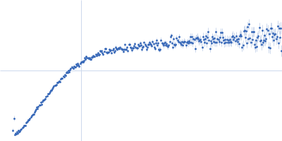
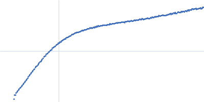
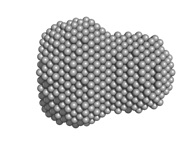
| Sample: |
L-lactate dehydrogenase tetramer, 141 kDa Plasmodium falciparum protein
|
| Buffer: |
100 mM Na-phosphate buffer, 400 mM NaCl, pH: 7.4
|
| Experiment: |
SAXS
data collected at Xenocs Xeuss 2.0 with MetalJet, Department of Macromolecular Physics, Faculty of Physics, Adam Mickiewicz University on 2019 Jul 3
|
A fragment-based approach identifies an allosteric pocket that impacts malate dehydrogenase activity
Communications Biology 4(1) (2021)
Reyes Romero A, Lunev S, Popowicz G, Calderone V, Gentili M, Sattler M, Plewka J, Taube M, Kozak M, Holak T, Dömling A, Groves M
|
| RgGuinier |
3.4 |
nm |
| Dmax |
10.5 |
nm |
| VolumePorod |
244 |
nm3 |
|
|
|
|
|
|
|
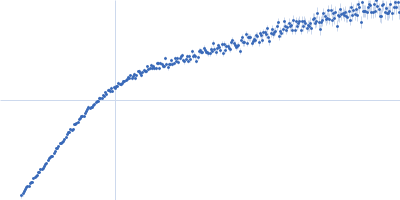
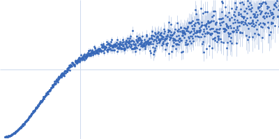
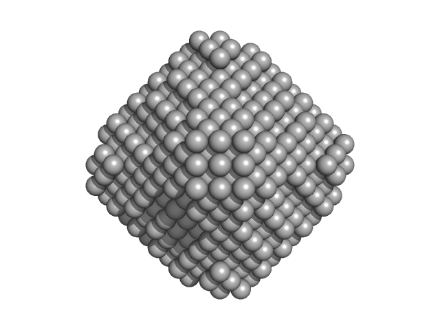
| Sample: |
L-lactate dehydrogenase tetramer, 141 kDa Plasmodium falciparum protein
|
| Buffer: |
100 mM Na-phosphate buffer, 400 mM NaCl, pH: 7.4
|
| Experiment: |
SAXS
data collected at Xenocs Xeuss 2.0 with MetalJet, Department of Macromolecular Physics, Faculty of Physics, Adam Mickiewicz University on 2019 Jul 3
|
A fragment-based approach identifies an allosteric pocket that impacts malate dehydrogenase activity
Communications Biology 4(1) (2021)
Reyes Romero A, Lunev S, Popowicz G, Calderone V, Gentili M, Sattler M, Plewka J, Taube M, Kozak M, Holak T, Dömling A, Groves M
|
| RgGuinier |
3.6 |
nm |
| Dmax |
11.4 |
nm |
| VolumePorod |
223 |
nm3 |
|
|
|
|
|
|
|
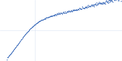
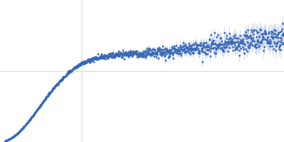
| Sample: |
Protein jagged-1 monomer, 115 kDa Mus musculus protein
|
| Buffer: |
20 mM HEPES, 150 mM NaCl, 2 mM CaCl2, pH: 7.4
|
| Experiment: |
SAXS
data collected at B21, Diamond Light Source on 2018 Dec 12
|
Notch-Jagged signaling complex defined by an interaction mosaic.
Proc Natl Acad Sci U S A 118(30) (2021)
Zeronian MR, Klykov O, Portell I de Montserrat J, Konijnenberg MJ, Gaur A, Scheltema RA, Janssen BJC
|
| RgGuinier |
7.4 |
nm |
| Dmax |
24.0 |
nm |
| VolumePorod |
375 |
nm3 |
|
|
|
|
|
|
|
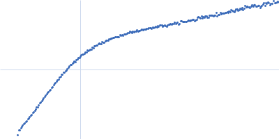
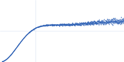
| Sample: |
Protein jagged-1 , 115 kDa Mus musculus protein
|
| Buffer: |
20 mM HEPES, 150 mM NaCl, 2 mM CaCl2, pH: 7.4
|
| Experiment: |
SAXS
data collected at B21, Diamond Light Source on 2018 Dec 11
|
Notch-Jagged signaling complex defined by an interaction mosaic.
Proc Natl Acad Sci U S A 118(30) (2021)
Zeronian MR, Klykov O, Portell I de Montserrat J, Konijnenberg MJ, Gaur A, Scheltema RA, Janssen BJC
|
| RgGuinier |
8.1 |
nm |
| Dmax |
30.0 |
nm |
| VolumePorod |
430 |
nm3 |
|
|
|
|
|
|
|
| Sample: |
Protein jagged-1 , 115 kDa Mus musculus protein
|
| Buffer: |
20 mM HEPES, 150 mM NaCl, 2 mM CaCl2, pH: 7.4
|
| Experiment: |
SAXS
data collected at B21, Diamond Light Source on 2018 Dec 11
|
Notch-Jagged signaling complex defined by an interaction mosaic.
Proc Natl Acad Sci U S A 118(30) (2021)
Zeronian MR, Klykov O, Portell I de Montserrat J, Konijnenberg MJ, Gaur A, Scheltema RA, Janssen BJC
|
| RgGuinier |
8.9 |
nm |
| Dmax |
33.0 |
nm |
| VolumePorod |
505 |
nm3 |
|
|
|
|
|
|
|
| Sample: |
Protein jagged-1 , 115 kDa Mus musculus protein
|
| Buffer: |
20 mM HEPES, 150 mM NaCl, 2 mM CaCl2, pH: 7.4
|
| Experiment: |
SAXS
data collected at B21, Diamond Light Source on 2018 Dec 11
|
Notch-Jagged signaling complex defined by an interaction mosaic.
Proc Natl Acad Sci U S A 118(30) (2021)
Zeronian MR, Klykov O, Portell I de Montserrat J, Konijnenberg MJ, Gaur A, Scheltema RA, Janssen BJC
|
| RgGuinier |
9.6 |
nm |
| Dmax |
42.0 |
nm |
| VolumePorod |
715 |
nm3 |
|
|
|
|
|
|
|
| Sample: |
Protein jagged-1 , 115 kDa Mus musculus protein
|
| Buffer: |
20 mM HEPES, 150 mM NaCl, 2 mM CaCl2, pH: 7.4
|
| Experiment: |
SAXS
data collected at B21, Diamond Light Source on 2018 Dec 11
|
Notch-Jagged signaling complex defined by an interaction mosaic.
Proc Natl Acad Sci U S A 118(30) (2021)
Zeronian MR, Klykov O, Portell I de Montserrat J, Konijnenberg MJ, Gaur A, Scheltema RA, Janssen BJC
|
| RgGuinier |
10.2 |
nm |
| Dmax |
43.0 |
nm |
| VolumePorod |
900 |
nm3 |
|
|
|
|
|
|
|
| Sample: |
Protein jagged-1 EGF8-11 , 21 kDa Mus musculus protein
|
| Buffer: |
20 mM HEPES, 150 mM NaCl, 2 mM CaCl2, pH: 7.4
|
| Experiment: |
SAXS
data collected at B21, Diamond Light Source on 2018 Dec 11
|
Notch-Jagged signaling complex defined by an interaction mosaic.
Proc Natl Acad Sci U S A 118(30) (2021)
Zeronian MR, Klykov O, Portell I de Montserrat J, Konijnenberg MJ, Gaur A, Scheltema RA, Janssen BJC
|
| RgGuinier |
3.3 |
nm |
| Dmax |
11.0 |
nm |
| VolumePorod |
26 |
nm3 |
|
|
|
|
|
|
|
| Sample: |
Protein jagged-1 EGF8-11 , 21 kDa Mus musculus protein
|
| Buffer: |
20 mM HEPES, 150 mM NaCl, 2 mM CaCl2, pH: 7.4
|
| Experiment: |
SAXS
data collected at B21, Diamond Light Source on 2018 Dec 11
|
Notch-Jagged signaling complex defined by an interaction mosaic.
Proc Natl Acad Sci U S A 118(30) (2021)
Zeronian MR, Klykov O, Portell I de Montserrat J, Konijnenberg MJ, Gaur A, Scheltema RA, Janssen BJC
|
| RgGuinier |
3.2 |
nm |
| Dmax |
11.5 |
nm |
| VolumePorod |
31 |
nm3 |
|
|
|
|
|
|
|
| Sample: |
Protein jagged-1 EGF8-11 , 21 kDa Mus musculus protein
|
| Buffer: |
20 mM HEPES, 150 mM NaCl, 2 mM CaCl2, pH: 7.4
|
| Experiment: |
SAXS
data collected at B21, Diamond Light Source on 2018 Dec 11
|
Notch-Jagged signaling complex defined by an interaction mosaic.
Proc Natl Acad Sci U S A 118(30) (2021)
Zeronian MR, Klykov O, Portell I de Montserrat J, Konijnenberg MJ, Gaur A, Scheltema RA, Janssen BJC
|
| RgGuinier |
3.1 |
nm |
| Dmax |
11.5 |
nm |
| VolumePorod |
34 |
nm3 |
|
|