|
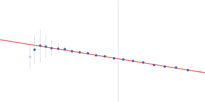
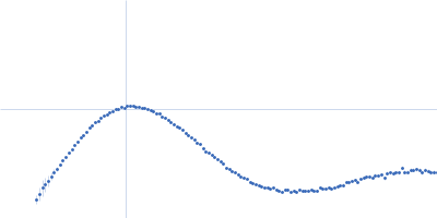
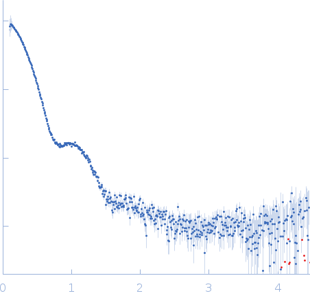
Synchrotron SAXS data from solutions of Zea mays beta-amylase (ZmBAM7) short in 50 mM HEPES, 25 mM NaCl, and 0.2 mM TCEP, pH 7.5 were collected on the 12.3.1 (SIBYLS) beam line at the Advanced Light Source (ALS; Berkeley, CA, USA) using a Pilatus3 X 2M detector at a sample-detector distance of 2 m and at a wavelength of λ = 0.127 nm (I(s) vs s, where s = 4πsinθ/λ, and 2θ is the scattering angle).
DNA sequences of ZmBAM7 (NP_001337631.1) were obtained from NCBI (https://www.ncbi.nlm.nih.gov/). The sequence of ZmBAM7-S was determined based on its in-frame start codon prediction 42 bases 5’ to the start of exon 2. The lengths of the predicted chloroplast transit peptides were determined using TargetP-2.0 (http://www.cbs.dtu.dk/services/TargetP/; Armenteros et al., 2019). Cell cultures of BL21 the ZmBAM7-S plasmid was grown to an optical density of 0.7 at 600 nm in Luria-Bertani media with 100 μg mL−1 carbenicillin at 37 °C and with shaking at 250 rpm. Isopropyl β-D-1-thiogalactopyranoside was added to a final concentration of 0.8 mM, and the flasks were shaken at 250 rpm at 20 °C overnight. Cells were pelleted by centrifugation at 3,000 rcf for 15 minutes at 4 °C then frozen at -80 °C for at least 10 minutes. Cell pellets were thawed and re-suspended in 50 mM NaH2PO4, 0.5 M NaCl, 0.2 mM tris(2-carboxyethyl)phosphine (TCEP), and 2 mM imidazole (pH 8.0) with EDTA-free protease inhibitor tablets (Pierce A32965). Cell lysis was completed by sonication in an ice bath (Misonix S-4000; Microtip) for 2.5 minutes at 55% amplitude (5 seconds on; 20 seconds off). After centrifugation of the cell lysate at 17,418 rcf for 20 minutes at 4 °C, the supernatants of AtBAM2 and ZmBAM7-S were separately loaded onto a TALON cobalt affinity column using an ÄKTA Start system (Cytiva Life Science, Marlborough, MA). Bound proteins were eluted from the TALON cobalt column using a step-wise addition of a second buffer containing 50 mM NaH2PO4, 0.5 M NaCl, 0.2 mM TCEP, and 200 mM imidazole (pH 8.0). The four 10 mL elution steps contained 12.5, 50, 125, or 200 mM imidazole mixed by the ÄKTA Start system. Fractions were analyzed for purity by SDS-PAGE using BioRad Mini-PROTEAN TGX Stain-Free gels in a buffer containing 250 mM Tris base, 1.92 M glycine, and 1% w/v SDS. Precision Plus Protein Unstained Standards (BioRad) were used as a marker for protein size. Dialysis using SpectraPor tubing (Spectrum, New Brunswick, NJ) with a molecular weight cutoff of 6-8 kD was completed overnight at 4 °C with constant stirring in 2 L of a buffer containing 20 mM HEPES, 100 mM NaCl, and 0.2 mM TCEP. The dialyzed proteins were concentrated in an Amicon Ultra-15 concentrator with a molecular weight cutoff of 10 kD at intervals of 30 minutes at 5,000 rcf and 4 °C until the desired volume was reached (~1.2 mL concentrated from 50 mL). Concentrated ZmBAM7-S was further purified using a HiLoad 16/60 Superdex 200 column (Cytiva) equilibrated with SEC buffer (50 mM HEPES, 25 mM NaCl, and 0.2 mM TCEP (pH 7.5)). Pure ZmBAM7-S, confirmed by SDS-PAGE, was concentrated as before, distributed into the plate for SAXS, and then flash frozen in liquid nitrogen. The concentration of ZmBAM7-S was determined using both the Bio-Rad Protein Assay Kit with BSA as the standard and by the absorbance at 280 nm using an extinction coefficient of 101,760 M-1cm-1. This value was calculated from the recombinant protein sequence including the His-tag using ProtParam (Gasteiger et al., 2005). Full length ZmBAM7-S was prepared for SAXS by diluting the SEC-purified protein using SEC buffer to five different concentrations (1.76, 2.64, 3.53, 5.29, and 6.17 mg mL-1) in 35 μL. Samples in a 96-well sample plate were flash frozen with liquid nitrogen. Three protein-free controls consisting of SEC buffer alone were included with the samples of both proteins. The sample plate was shipped overnight on dry ice to the Advanced Light Source at Lawrence Berkeley National Laboratory. Prior to data collection (date of collection: 12/07/2020), the plate was spun at 3700 rev min-1 for 10 minutes by beam line staff. Scattering data on the samples and controls were collected every 0.3 seconds for a total of 10 seconds resulting in 33 frames of data per sample. The beam energy was 11 keV, and the detector was 2 meters from the sample holder. Samples were kept at 10 °C during collection. Data from the protein free buffer was collected before and after ZmBAM7-S and truncated ZmBAM7-S sample collection to ensure there was no difference in scattering due to contamination of the sample cell. Buffer scattering was subtracted from sample scattering in RAW (version 2.0.3) using SAXS FrameSlice (version 1.4.13) as a guide to determine which frames were used during buffer subtraction (Hopkins et al., 2017).
|
|
 s, nm-1