Search

 
Advanced search  

37 hits found for Korasick

SASDCP3 – Proline utilization A from Bdellovibrio bacteriovorus

Bifunctional protein PutA experimental SAS data
DAMMIF model
Sample: 
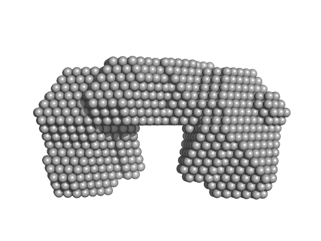
Bifunctional protein PutA dimer, 219 kDa Bdellovibrio bacteriovorus protein
Buffer: 50 mM Tris, 125 mM NaCl, 1 mM EDTA, and 1 mM tris(3-hydroxypropyl)phosphine (THP) at pH 7.5,, pH: 7.5
Experiment: SAXS data collected at 12.3.1 (SIBYLS), Advanced Light Source (ALS) on 2012 Jun 8
Biophysical investigation of type A PutAs reveals a conserved core oligomeric structure. FEBS J 284(18):3029-3049 (2017)
Korasick DA, Singh H, Pemberton TA, Luo M, Dhatwalia R, Tanner JJ
RgGuinier 4.5 nm
Dmax 14.0 nm
VolumePorod 287 nm3

SASDDP3 – N-propargyl glycine-Inactivated Proline utilization A from Bradyrhizobium diazoefficiens (formerly Bradyrhizobium japonicum) collected by SEC-SAXS

Bifunctional protein PutA experimental SAS data
PDB (PROTEIN DATA BANK) model
Sample: Bifunctional protein PutA dimer, 215 kDa Bradyrhizobium diazoefficiens protein
Buffer: 50 mM Tris, 50 mM NaCl, 0.5 mM TCEP, 5% (v/v) glycerol, pH: 7.8
Experiment: SAXS data collected at BioCAT 18ID, Advanced Photon Source (APS), Argonne National Laboratory on 2017 Jul 16
Redox Modulation of Oligomeric State in Proline Utilization A. Biophys J 114(12):2833-2843 (2018)
Korasick DA, Campbell AC, Christgen SL, Chakravarthy S, White TA, Becker DF, Tanner JJ
RgGuinier 4.6 nm
Dmax 14.4 nm
VolumePorod 324 nm3

SASDCQ3 – Proline utilization A from Desulfovibrio vulgaris 1.5 mg/mL

Bifunctional protein PutA experimental SAS data
Bifunctional protein PutA Kratky plot
Sample: Bifunctional protein PutA dimer, 229 kDa Desulfovibrio vulgaris protein
Buffer: 50 mM Tris-HCl, 50 mM NaCl, 0.5 mM EDTA, and 0.5 mM THP at pH 7.5., pH: 7.5
Experiment: SAXS data collected at 12.3.1 (SIBYLS), Advanced Light Source (ALS) on 2012 Jun 8
Biophysical investigation of type A PutAs reveals a conserved core oligomeric structure. FEBS J 284(18):3029-3049 (2017)
Korasick DA, Singh H, Pemberton TA, Luo M, Dhatwalia R, Tanner JJ
RgGuinier 4.4 nm
Dmax 16.0 nm
VolumePorod 293 nm3

SASDDQ3 – Proline utilization A from Bradyrhizobium diazoefficiens (formerly Bradyrhizobium japonicum) collected by SEC-SAXS

Bifunctional protein PutA experimental SAS data
PDB (PROTEIN DATA BANK) model
Sample: Bifunctional protein PutA tetramer, 430 kDa Bradyrhizobium diazoefficiens protein
Buffer: 50 mM Tris, 50 mM NaCl, 0.5 mM TCEP, 5% (v/v) glycerol, pH: 7.8
Experiment: SAXS data collected at BioCAT 18ID, Advanced Photon Source (APS), Argonne National Laboratory on 2017 Jul 16
Redox Modulation of Oligomeric State in Proline Utilization A. Biophys J 114(12):2833-2843 (2018)
Korasick DA, Campbell AC, Christgen SL, Chakravarthy S, White TA, Becker DF, Tanner JJ
RgGuinier 5.2 nm
Dmax 14.2 nm
VolumePorod 582 nm3

SASDCR3 – Proline utilization A from Legionella pneumophila 3 mg/mL

Bifunctional protein PutA experimental SAS data
Bifunctional protein PutA Kratky plot
Sample: Bifunctional protein PutA dimer, 238 kDa Legionella pneumophila subsp. … protein
Buffer: 50 mM Tris-HCl, 50 mM NaCl, 0.5 mM EDTA, and 0.5 mM THP at pH 7.5., pH: 7.5
Experiment: SAXS data collected at 12.3.1 (SIBYLS), Advanced Light Source (ALS) on 2010 Apr 20
Biophysical investigation of type A PutAs reveals a conserved core oligomeric structure. FEBS J 284(18):3029-3049 (2017)
Korasick DA, Singh H, Pemberton TA, Luo M, Dhatwalia R, Tanner JJ
RgGuinier 4.6 nm
Dmax 16.0 nm
VolumePorod 291 nm3

SASDCS3 – Proline utilization A from Bradyrhizobium diazoefficiens (formerly Bradyrhizobium japonicum) 2.3 mg/mL

Proline dehydrogenase experimental SAS data
Proline dehydrogenase Kratky plot
Sample: Proline dehydrogenase tetramer, 430 kDa Bradyrhizobium diazoefficiens protein
Buffer: 50 mM Tris (pH 7.8), 50 mM NaCl, 0.5 mM Tris(2-carboxyethyl)phosphine, and 5% (v/v) glycerol, pH: 7.8
Experiment: SAXS data collected at 12.3.1 (SIBYLS), Advanced Light Source (ALS) on 2016 Dec 16
Biophysical investigation of type A PutAs reveals a conserved core oligomeric structure. FEBS J 284(18):3029-3049 (2017)
Korasick DA, Singh H, Pemberton TA, Luo M, Dhatwalia R, Tanner JJ
RgGuinier 5.3 nm
Dmax 14.1 nm
VolumePorod 541 nm3

SASDCT3 – Proline utilization A from Bradyrhizobium diazoefficiens (formerly Bradyrhizobium japonicum) 4.7 mg/mL

Proline dehydrogenase experimental SAS data
Proline dehydrogenase Kratky plot
Sample: Proline dehydrogenase tetramer, 430 kDa Bradyrhizobium diazoefficiens protein
Buffer: 50 mM Tris (pH 7.8), 50 mM NaCl, 0.5 mM Tris(2-carboxyethyl)phosphine, and 5% (v/v) glycerol, pH: 7.8
Experiment: SAXS data collected at 12.3.1 (SIBYLS), Advanced Light Source (ALS) on 2016 Dec 12
Biophysical investigation of type A PutAs reveals a conserved core oligomeric structure. FEBS J 284(18):3029-3049 (2017)
Korasick DA, Singh H, Pemberton TA, Luo M, Dhatwalia R, Tanner JJ
RgGuinier 5.2 nm
Dmax 14.6 nm
VolumePorod 553 nm3

SASDCU3 – Proline utilization A from Bradyrhizobium diazoefficiens (formerly Bradyrhizobium japonicum) 7.0 mg/mL

Proline dehydrogenase experimental SAS data
DAMMIF model
Sample: Proline dehydrogenase tetramer, 430 kDa Bradyrhizobium diazoefficiens protein
Buffer: 50 mM Tris (pH 7.8), 50 mM NaCl, 0.5 mM Tris(2-carboxyethyl)phosphine, and 5% (v/v) glycerol, pH: 7.8
Experiment: SAXS data collected at 12.3.1 (SIBYLS), Advanced Light Source (ALS) on 2016 Dec 12
Biophysical investigation of type A PutAs reveals a conserved core oligomeric structure. FEBS J 284(18):3029-3049 (2017)
Korasick DA, Singh H, Pemberton TA, Luo M, Dhatwalia R, Tanner JJ
RgGuinier 5.2 nm
Dmax 13.7 nm
VolumePorod 560 nm3

SASDCV3 – Proline utilization A from Legionella pneumophila 5 mg/mL

Bifunctional protein PutA experimental SAS data
Bifunctional protein PutA Kratky plot
Sample: Bifunctional protein PutA dimer, 238 kDa Legionella pneumophila subsp. … protein
Buffer: 50 mM Tris-HCl, 50 mM NaCl, 0.5 mM EDTA, and 0.5 mM THP at pH 7.5., pH: 7.5
Experiment: SAXS data collected at 12.3.1 (SIBYLS), Advanced Light Source (ALS) on 2010 Apr 20
Biophysical investigation of type A PutAs reveals a conserved core oligomeric structure. FEBS J 284(18):3029-3049 (2017)
Korasick DA, Singh H, Pemberton TA, Luo M, Dhatwalia R, Tanner JJ
RgGuinier 4.6 nm
Dmax 15.3 nm
VolumePorod 297 nm3

SASDCW3 – Proline utilization A from Legionella pneumophila 8 mg/mL

Bifunctional protein PutA experimental SAS data
DAMMIF model
Sample: Bifunctional protein PutA dimer, 238 kDa Legionella pneumophila subsp. … protein
Buffer: 50 mM Tris-HCl, 50 mM NaCl, 0.5 mM EDTA, and 0.5 mM THP at pH 7.5., pH: 7.5
Experiment: SAXS data collected at 12.3.1 (SIBYLS), Advanced Light Source (ALS) on 2010 Apr 20
Biophysical investigation of type A PutAs reveals a conserved core oligomeric structure. FEBS J 284(18):3029-3049 (2017)
Korasick DA, Singh H, Pemberton TA, Luo M, Dhatwalia R, Tanner JJ
RgGuinier 4.6 nm
Dmax 15.5 nm
VolumePorod 295 nm3

SASDCX3 – Proline utilization A from Desulfovibrio vulgaris 3.0 mg/mL

Bifunctional protein PutA experimental SAS data
DAMMIF model
Sample: Bifunctional protein PutA dimer, 229 kDa Desulfovibrio vulgaris protein
Buffer: 50 mM Tris-HCl, 50 mM NaCl, 0.5 mM EDTA, and 0.5 mM THP at pH 7.5., pH: 7.5
Experiment: SAXS data collected at 12.3.1 (SIBYLS), Advanced Light Source (ALS) on 2012 Jun 8
Biophysical investigation of type A PutAs reveals a conserved core oligomeric structure. FEBS J 284(18):3029-3049 (2017)
Korasick DA, Singh H, Pemberton TA, Luo M, Dhatwalia R, Tanner JJ
RgGuinier 4.4 nm
Dmax 16.0 nm
VolumePorod 295 nm3

SASDCY3 – Proline utilization A from Desulfovibrio vulgaris 4.5 mg/mL

Bifunctional protein PutA experimental SAS data
Bifunctional protein PutA Kratky plot
Sample: Bifunctional protein PutA dimer, 229 kDa Desulfovibrio vulgaris protein
Buffer: 50 mM Tris-HCl, 50 mM NaCl, 0.5 mM EDTA, and 0.5 mM THP at pH 7.5., pH: 7.5
Experiment: SAXS data collected at 12.3.1 (SIBYLS), Advanced Light Source (ALS) on 2012 Jun 8
Biophysical investigation of type A PutAs reveals a conserved core oligomeric structure. FEBS J 284(18):3029-3049 (2017)
Korasick DA, Singh H, Pemberton TA, Luo M, Dhatwalia R, Tanner JJ
RgGuinier 4.4 nm
Dmax 16.0 nm
VolumePorod 294 nm3

SASDCZ3 – Proline utilization A from Bradyrhizobium diazoefficiens (formerly Bradyrhizobium japonicum) R51E mutant 2.3 mg/mL

Proline dehydrogenase experimental SAS data
Proline dehydrogenase Kratky plot
Sample: Proline dehydrogenase dimer, 215 kDa Bradyrhizobium diazoefficiens protein
Buffer: 50 mM Tris (pH 7.8), 50 mM NaCl, 0.5 mM Tris(2-carboxyethyl)phosphine, and 5% (v/v) glycerol, pH: 7.8
Experiment: SAXS data collected at 12.3.1 (SIBYLS), Advanced Light Source (ALS) on 2016 Dec 16
Biophysical investigation of type A PutAs reveals a conserved core oligomeric structure. FEBS J 284(18):3029-3049 (2017)
Korasick DA, Singh H, Pemberton TA, Luo M, Dhatwalia R, Tanner JJ
RgGuinier 4.5 nm
Dmax 14.5 nm
VolumePorod 281 nm3

SASDC24 – Proline utilization A from Bradyrhizobium diazoefficiens (formerly Bradyrhizobium japonicum) R51E mutant 4.7 mg/mL

Proline dehydrogenase experimental SAS data
Proline dehydrogenase Kratky plot
Sample: Proline dehydrogenase dimer, 215 kDa Bradyrhizobium diazoefficiens protein
Buffer: 50 mM Tris (pH 7.8), 50 mM NaCl, 0.5 mM Tris(2-carboxyethyl)phosphine, and 5% (v/v) glycerol, pH: 7.8
Experiment: SAXS data collected at 12.3.1 (SIBYLS), Advanced Light Source (ALS) on 2016 Dec 16
Biophysical investigation of type A PutAs reveals a conserved core oligomeric structure. FEBS J 284(18):3029-3049 (2017)
Korasick DA, Singh H, Pemberton TA, Luo M, Dhatwalia R, Tanner JJ
RgGuinier 4.5 nm
Dmax 13.9 nm
VolumePorod 283 nm3

SASDC34 – Proline utilization A from Bradyrhizobium diazoefficiens (formerly Bradyrhizobium japonicum) R51E mutant 7.0 mg/mL

Proline dehydrogenase experimental SAS data
DAMMIF model
Sample: Proline dehydrogenase dimer, 215 kDa Bradyrhizobium diazoefficiens protein
Buffer: 50 mM Tris (pH 7.8), 50 mM NaCl, 0.5 mM Tris(2-carboxyethyl)phosphine, and 5% (v/v) glycerol, pH: 7.8
Experiment: SAXS data collected at 12.3.1 (SIBYLS), Advanced Light Source (ALS) on 2016 Dec 16
Biophysical investigation of type A PutAs reveals a conserved core oligomeric structure. FEBS J 284(18):3029-3049 (2017)
Korasick DA, Singh H, Pemberton TA, Luo M, Dhatwalia R, Tanner JJ
RgGuinier 4.5 nm
Dmax 14.6 nm
VolumePorod 289 nm3

SASDGH4 – Human alpha-aminoadipic semialdehyde dehydrogenase (ALDH)7A1 at 1.2 mg/mL

Alpha-aminoadipic semialdehyde dehydrogenase experimental SAS data
PDB (PROTEIN DATA BANK) model
Sample: Alpha-aminoadipic semialdehyde dehydrogenase tetramer, 222 kDa Homo sapiens protein
Buffer: 50 mM HEPES, 100 mM NaCl, 1 mM DTT, 10 mM NAD, 2% (v/v) glycerol, pH: 8
Experiment: SAXS data collected at 12.3.1 (SIBYLS), Advanced Light Source (ALS) on 2019 May 1
Structural Analysis of Pathogenic Mutations Targeting Glu427 of ALDH7A1, the Hot Spot Residue of Pyridoxine-Dependent Epilepsy. J Inherit Metab Dis (2019)
...Korasick DA, Gates KS, Tanner JJ
RgGuinier 3.8 nm
Dmax 11.5 nm
VolumePorod 350 nm3

SASDGJ4 – Human alpha-aminoadipic semialdehyde dehydrogenase (ALDH)7A1 at 2.3 mg/mL

Alpha-aminoadipic semialdehyde dehydrogenase experimental SAS data
PDB (PROTEIN DATA BANK) model
Sample: Alpha-aminoadipic semialdehyde dehydrogenase tetramer, 222 kDa Homo sapiens protein
Buffer: 50 mM HEPES, 100 mM NaCl, 1 mM DTT, 10 mM NAD, 2% (v/v) glycerol, pH: 8
Experiment: SAXS data collected at 12.3.1 (SIBYLS), Advanced Light Source (ALS) on 2019 May 1
Structural Analysis of Pathogenic Mutations Targeting Glu427 of ALDH7A1, the Hot Spot Residue of Pyridoxine-Dependent Epilepsy. J Inherit Metab Dis (2019)
...Korasick DA, Gates KS, Tanner JJ
RgGuinier 3.8 nm
Dmax 10.7 nm
VolumePorod 326 nm3

SASDGK4 – Human alpha-aminoadipic semialdehyde dehydrogenase (ALDH)7A1 at 4.7 mg/mL

Alpha-aminoadipic semialdehyde dehydrogenase experimental SAS data
PDB (PROTEIN DATA BANK) model
Sample: Alpha-aminoadipic semialdehyde dehydrogenase tetramer, 222 kDa Homo sapiens protein
Buffer: 50 mM HEPES, 100 mM NaCl, 1 mM DTT, 10 mM NAD, 2% (v/v) glycerol, pH: 8
Experiment: SAXS data collected at 12.3.1 (SIBYLS), Advanced Light Source (ALS) on 2019 May 1
Structural Analysis of Pathogenic Mutations Targeting Glu427 of ALDH7A1, the Hot Spot Residue of Pyridoxine-Dependent Epilepsy. J Inherit Metab Dis (2019)
...Korasick DA, Gates KS, Tanner JJ
RgGuinier 3.8 nm
Dmax 10.7 nm
VolumePorod 315 nm3

SASDGL4 – Human alpha-aminoadipic semialdehyde dehydrogenase (ALDH)7A1 E399Q at 1.1 mg/mL

Alpha-aminoadipic semialdehyde dehydrogenase E399Q experimental SAS data
PDB (PROTEIN DATA BANK) model
Sample: Alpha-aminoadipic semialdehyde dehydrogenase E399Q , 56 kDa Homo sapiens protein
Buffer: 50 mM HEPES, 100 mM NaCl, 1 mM DTT, 10 mM NAD, 2% (v/v) glycerol, pH: 8
Experiment: SAXS data collected at 12.3.1 (SIBYLS), Advanced Light Source (ALS) on 2019 May 1
Structural Analysis of Pathogenic Mutations Targeting Glu427 of ALDH7A1, the Hot Spot Residue of Pyridoxine-Dependent Epilepsy. J Inherit Metab Dis (2019)
...Korasick DA, Gates KS, Tanner JJ
RgGuinier 3.7 nm
Dmax 10.3 nm
VolumePorod 237 nm3

SASDGM4 – Human alpha-aminoadipic semialdehyde dehydrogenase (ALDH)7A1 E399Q at 2.1 mg/mL

Alpha-aminoadipic semialdehyde dehydrogenase E399Q experimental SAS data
PDB (PROTEIN DATA BANK) model
Sample: Alpha-aminoadipic semialdehyde dehydrogenase E399Q , 56 kDa Homo sapiens protein
Buffer: 50 mM HEPES, 100 mM NaCl, 1 mM DTT, 10 mM NAD, 2% (v/v) glycerol, pH: 8
Experiment: SAXS data collected at 12.3.1 (SIBYLS), Advanced Light Source (ALS) on 2019 May 1
Structural Analysis of Pathogenic Mutations Targeting Glu427 of ALDH7A1, the Hot Spot Residue of Pyridoxine-Dependent Epilepsy. J Inherit Metab Dis (2019)
...Korasick DA, Gates KS, Tanner JJ
RgGuinier 3.8 nm
Dmax 10.8 nm
VolumePorod 238 nm3

SASDGN4 – Human alpha-aminoadipic semialdehyde dehydrogenase (ALDH)7A1 E399Q at 4.3 mg/mL

Alpha-aminoadipic semialdehyde dehydrogenase E399Q experimental SAS data
PDB (PROTEIN DATA BANK) model
Sample: Alpha-aminoadipic semialdehyde dehydrogenase E399Q , 56 kDa Homo sapiens protein
Buffer: 50 mM HEPES, 100 mM NaCl, 1 mM DTT, 10 mM NAD, 2% (v/v) glycerol, pH: 8
Experiment: SAXS data collected at 12.3.1 (SIBYLS), Advanced Light Source (ALS) on 2019 May 1
Structural Analysis of Pathogenic Mutations Targeting Glu427 of ALDH7A1, the Hot Spot Residue of Pyridoxine-Dependent Epilepsy. J Inherit Metab Dis (2019)
...Korasick DA, Gates KS, Tanner JJ
RgGuinier 3.8 nm
Dmax 10.6 nm
VolumePorod 255 nm3

SASDGP4 – Human alpha-aminoadipic semialdehyde dehydrogenase (ALDH)7A1 E399D at 1.4 mg/mL

Alpha-aminoadipic semialdehyde dehydrogenase experimental SAS data
PDB (PROTEIN DATA BANK) model
Sample: Alpha-aminoadipic semialdehyde dehydrogenase , 56 kDa Homo sapiens protein
Buffer: 50 mM HEPES, 100 mM NaCl, 1 mM DTT, 10 mM NAD, 2% (v/v) glycerol, pH: 8
Experiment: SAXS data collected at 12.3.1 (SIBYLS), Advanced Light Source (ALS) on 2019 May 1
Structural Analysis of Pathogenic Mutations Targeting Glu427 of ALDH7A1, the Hot Spot Residue of Pyridoxine-Dependent Epilepsy. J Inherit Metab Dis (2019)
...Korasick DA, Gates KS, Tanner JJ
RgGuinier 3.7 nm
Dmax 11.3 nm
VolumePorod 256 nm3

SASDGQ4 – Human alpha-aminoadipic semialdehyde dehydrogenase (ALDH)7A1 E399D at 2.9 mg/mL

Alpha-aminoadipic semialdehyde dehydrogenase experimental SAS data
PDB (PROTEIN DATA BANK) model
Sample: Alpha-aminoadipic semialdehyde dehydrogenase , 56 kDa Homo sapiens protein
Buffer: 50 mM HEPES, 100 mM NaCl, 1 mM DTT, 10 mM NAD, 2% (v/v) glycerol, pH: 8
Experiment: SAXS data collected at 12.3.1 (SIBYLS), Advanced Light Source (ALS) on 2019 May 1
Structural Analysis of Pathogenic Mutations Targeting Glu427 of ALDH7A1, the Hot Spot Residue of Pyridoxine-Dependent Epilepsy. J Inherit Metab Dis (2019)
...Korasick DA, Gates KS, Tanner JJ
RgGuinier 3.8 nm
Dmax 11.4 nm
VolumePorod 260 nm3

SASDGR4 – Human alpha-aminoadipic semialdehyde dehydrogenase (ALDH)7A1 E399D at 5.7 mg/mL

Alpha-aminoadipic semialdehyde dehydrogenase E399D experimental SAS data
PDB (PROTEIN DATA BANK) model
Sample: Alpha-aminoadipic semialdehyde dehydrogenase E399D , 56 kDa Homo sapiens protein
Buffer: 50 mM HEPES, 100 mM NaCl, 1 mM DTT, 10 mM NAD, 2% (v/v) glycerol, pH: 8
Experiment: SAXS data collected at 12.3.1 (SIBYLS), Advanced Light Source (ALS) on 2019 May 1
Structural Analysis of Pathogenic Mutations Targeting Glu427 of ALDH7A1, the Hot Spot Residue of Pyridoxine-Dependent Epilepsy. J Inherit Metab Dis (2019)
...Korasick DA, Gates KS, Tanner JJ
RgGuinier 3.9 nm
Dmax 10.6 nm
VolumePorod 272 nm3

SASDGS4 – Human alpha-aminoadipic semialdehyde dehydrogenase (ALDH)7A1 E399G at 1.6 mg/mL

Alpha-aminoadipic semialdehyde dehydrogenase E399G experimental SAS data
PDB (PROTEIN DATA BANK) model
Sample: Alpha-aminoadipic semialdehyde dehydrogenase E399G , 55 kDa Homo sapiens protein
Buffer: 50 mM HEPES, 100 mM NaCl, 1 mM DTT, 10 mM NAD, 2% (v/v) glycerol, pH: 8
Experiment: SAXS data collected at 12.3.1 (SIBYLS), Advanced Light Source (ALS) on 2019 May 1
Structural Analysis of Pathogenic Mutations Targeting Glu427 of ALDH7A1, the Hot Spot Residue of Pyridoxine-Dependent Epilepsy. J Inherit Metab Dis (2019)
...Korasick DA, Gates KS, Tanner JJ
RgGuinier 3.8 nm
Dmax 11.3 nm
VolumePorod 290 nm3

SASDGT4 – Human alpha-aminoadipic semialdehyde dehydrogenase (ALDH)7A1 E399G at 3.2 mg/mL

Alpha-aminoadipic semialdehyde dehydrogenase E399G experimental SAS data
PDB (PROTEIN DATA BANK) model
Sample: Alpha-aminoadipic semialdehyde dehydrogenase E399G , 55 kDa Homo sapiens protein
Buffer: 50 mM HEPES, 100 mM NaCl, 1 mM DTT, 10 mM NAD, 2% (v/v) glycerol, pH: 8
Experiment: SAXS data collected at 12.3.1 (SIBYLS), Advanced Light Source (ALS) on 2019 May 1
Structural Analysis of Pathogenic Mutations Targeting Glu427 of ALDH7A1, the Hot Spot Residue of Pyridoxine-Dependent Epilepsy. J Inherit Metab Dis (2019)
...Korasick DA, Gates KS, Tanner JJ
RgGuinier 3.7 nm
Dmax 10.6 nm
VolumePorod 250 nm3

SASDGU4 – Human alpha-aminoadipic semialdehyde dehydrogenase (ALDH)7A1 E399G at 6.5 mg/mL

Alpha-aminoadipic semialdehyde dehydrogenase E399G experimental SAS data
PDB (PROTEIN DATA BANK) model
Sample: Alpha-aminoadipic semialdehyde dehydrogenase E399G , 55 kDa Homo sapiens protein
Buffer: 50 mM HEPES, 100 mM NaCl, 1 mM DTT, 10 mM NAD, 2% (v/v) glycerol, pH: 8
Experiment: SAXS data collected at 12.3.1 (SIBYLS), Advanced Light Source (ALS) on 2019 May 1
Structural Analysis of Pathogenic Mutations Targeting Glu427 of ALDH7A1, the Hot Spot Residue of Pyridoxine-Dependent Epilepsy. J Inherit Metab Dis (2019)
...Korasick DA, Gates KS, Tanner JJ
RgGuinier 3.8 nm
Dmax 10.0 nm
VolumePorod 270 nm3

SASDHV5 – Human 4-trimethylaminobutyraldehyde dehydrogenase (ALDH9A1) at 1.25 mg/mL

4-trimethylaminobutyraldehyde dehydrogenase experimental SAS data
OTHER model
Sample: 4-trimethylaminobutyraldehyde dehydrogenase tetramer, 215 kDa Homo sapiens protein
Buffer: 50 mM HEPES, 600 mM NaCl, 2% (v/v) glycerol, 1 mM DTT, 1 mM NAD+, pH: 8
Experiment: SAXS data collected at 12.3.1 (SIBYLS), Advanced Light Source (ALS) on 2019 May 28
Inhibition, crystal structures, and in-solution oligomeric structure of aldehyde dehydrogenase 9A1. Arch Biochem Biophys :108477 (2020)
...Korasick DA, Qureshi IA, Campbell AC, Gates KS, Tanner JJ
RgGuinier 3.8 nm
Dmax 10.9 nm
VolumePorod 252 nm3

SASDHW5 – Human 4-trimethylaminobutyraldehyde dehydrogenase (ALDH9A1) at 2.5 mg/mL

4-trimethylaminobutyraldehyde dehydrogenase experimental SAS data
OTHER model
Sample: 4-trimethylaminobutyraldehyde dehydrogenase tetramer, 215 kDa Homo sapiens protein
Buffer: 50 mM HEPES, 600 mM NaCl, 2% (v/v) glycerol, 1 mM DTT, 1 mM NAD+, pH: 8
Experiment: SAXS data collected at 12.3.1 (SIBYLS), Advanced Light Source (ALS) on 2019 May 28
Inhibition, crystal structures, and in-solution oligomeric structure of aldehyde dehydrogenase 9A1. Arch Biochem Biophys :108477 (2020)
...Korasick DA, Qureshi IA, Campbell AC, Gates KS, Tanner JJ
RgGuinier 3.8 nm
Dmax 10.8 nm
VolumePorod 240 nm3

SASDHX5 – Human 4-trimethylaminobutyraldehyde dehydrogenase (ALDH9A1) at 5 mg/mL

4-trimethylaminobutyraldehyde dehydrogenase experimental SAS data
OTHER model
Sample: 4-trimethylaminobutyraldehyde dehydrogenase tetramer, 215 kDa Homo sapiens protein
Buffer: 50 mM HEPES, 600 mM NaCl, 2% (v/v) glycerol, 1 mM DTT, 1 mM NAD+, pH: 8
Experiment: SAXS data collected at 12.3.1 (SIBYLS), Advanced Light Source (ALS) on 2019 May 28
Inhibition, crystal structures, and in-solution oligomeric structure of aldehyde dehydrogenase 9A1. Arch Biochem Biophys :108477 (2020)
...Korasick DA, Qureshi IA, Campbell AC, Gates KS, Tanner JJ
RgGuinier 3.8 nm
Dmax 10.5 nm
VolumePorod 240 nm3

SASDE96 – Aldehyde dehydrogenase 12 from Zea mays Extrapolated to Infinite Dilution

Aldehyde dehydrogenase 12 experimental SAS data
ALLOSMOD model
Sample: Aldehyde dehydrogenase 12 tetramer, 242 kDa Zea mays protein
Buffer: 50 mM Tris-HCl, 50 mM NaCl, 0.5 mM TCEP, and 5% (v/v) glycerol, pH: 7.8
Experiment: SAXS data collected at 12.3.1 (SIBYLS), Advanced Light Source (ALS) on 2016 Dec 6
Structural and Biochemical Characterization of Aldehyde Dehydrogenase 12, the Last Enzyme of Proline Catabolism in Plants. J Mol Biol (2018)
Korasick DA, Končitíková R, Kopečná M, Hájková E, Vigouroux A, Moréra S, Becker DF, Šebela M, Tanner JJ, Kopečný D
RgGuinier 4.1 nm
Dmax 14.4 nm
VolumePorod 351 nm3

SASDD29 – Low load concentration of apo alpha-aminoadipic semialdehyde dehydrogenase (ALDH7A1) collected by SEC-SAXS

Alpha-aminoadipic semialdehyde dehydrogenase experimental SAS data
PDB (PROTEIN DATA BANK) model
Sample: Alpha-aminoadipic semialdehyde dehydrogenase tetramer, 222 kDa Homo sapiens protein
Buffer: 50 mM Tris, 50 mM NaCl, 0.5 mM DTT, 5% (v/v) glycerol, pH: 7.8
Experiment: SAXS data collected at BioCAT 18ID, Advanced Photon Source (APS), Argonne National Laboratory on 2018 Feb 22
NAD+ Promotes Assembly of the Active Tetramer of Aldehyde Dehydrogenase 7A1. FEBS Lett (2018)
Korasick DA, White TA, Chakravarthy S, Tanner JJ
RgGuinier 3.5 nm
VolumePorod 212 nm3

SASDD39 – Medium load concentration of apo alpha-aminoadipic semialdehyde dehydrogenase (ALDH7A1) collected by SEC-SAXS

Alpha-aminoadipic semialdehyde dehydrogenase experimental SAS data
PDB (PROTEIN DATA BANK) model
Sample: Alpha-aminoadipic semialdehyde dehydrogenase tetramer, 222 kDa Homo sapiens protein
Buffer: 50 mM Tris, 50 mM NaCl, 0.5 mM DTT, 5% (v/v) glycerol, pH: 7.8
Experiment: SAXS data collected at BioCAT 18ID, Advanced Photon Source (APS), Argonne National Laboratory on 2018 Feb 22
NAD+ Promotes Assembly of the Active Tetramer of Aldehyde Dehydrogenase 7A1. FEBS Lett (2018)
Korasick DA, White TA, Chakravarthy S, Tanner JJ
RgGuinier 3.7 nm
VolumePorod 229 nm3

SASDD49 – High load concentration of apo alpha-aminoadipic semialdehyde dehydrogenase (ALDH7A1) collected by SEC-SAXS

Alpha-aminoadipic semialdehyde dehydrogenase experimental SAS data
PDB (PROTEIN DATA BANK) model
Sample: Alpha-aminoadipic semialdehyde dehydrogenase tetramer, 222 kDa Homo sapiens protein
Buffer: 50 mM Tris, 50 mM NaCl, 0.5 mM DTT, 5% (v/v) glycerol, pH: 7.8
Experiment: SAXS data collected at BioCAT 18ID, Advanced Photon Source (APS), Argonne National Laboratory on 2018 Feb 22
NAD+ Promotes Assembly of the Active Tetramer of Aldehyde Dehydrogenase 7A1. FEBS Lett (2018)
Korasick DA, White TA, Chakravarthy S, Tanner JJ
RgGuinier 3.7 nm
VolumePorod 238 nm3

SASDD59 – Low load concentration of alpha-aminoadipic semialdehyde dehydrogenase (ALDH7A1) with nicotinamide adenine dinucleotide (NAD) collected by SEC-SAXS

Alpha-aminoadipic semialdehyde dehydrogenase experimental SAS data
PDB (PROTEIN DATA BANK) model
Sample: Alpha-aminoadipic semialdehyde dehydrogenase tetramer, 222 kDa Homo sapiens protein
Buffer: 50 mM Tris, 50 mM NaCl, 0.5 mM DTT, 5% (v/v) glycerol, 1 mM NAD, pH: 7.8
Experiment: SAXS data collected at BioCAT 18ID, Advanced Photon Source (APS), Argonne National Laboratory on 2018 Feb 22
NAD+ Promotes Assembly of the Active Tetramer of Aldehyde Dehydrogenase 7A1. FEBS Lett (2018)
Korasick DA, White TA, Chakravarthy S, Tanner JJ
RgGuinier 3.7 nm
VolumePorod 277 nm3

SASDD69 – Medium load concentration of alpha-aminoadipic semialdehyde dehydrogenase (ALDH7A1) with nicotinamide adenine dinucleotide (NAD) collected by SEC-SAXS

Alpha-aminoadipic semialdehyde dehydrogenase experimental SAS data
PDB (PROTEIN DATA BANK) model
Sample: Alpha-aminoadipic semialdehyde dehydrogenase tetramer, 222 kDa Homo sapiens protein
Buffer: 50 mM Tris, 50 mM NaCl, 0.5 mM DTT, 5% (v/v) glycerol, 1 mM NAD, pH: 7.8
Experiment: SAXS data collected at BioCAT 18ID, Advanced Photon Source (APS), Argonne National Laboratory on 2018 Feb 22
NAD+ Promotes Assembly of the Active Tetramer of Aldehyde Dehydrogenase 7A1. FEBS Lett (2018)
Korasick DA, White TA, Chakravarthy S, Tanner JJ
RgGuinier 3.8 nm
VolumePorod 275 nm3

SASDD79 – High load concentration of alpha-aminoadipic semialdehyde dehydrogenase ALDH7A1 with nicotinamide adenine dinucleotide (NAD) collected by SEC-SAXS

Alpha-aminoadipic semialdehyde dehydrogenase experimental SAS data
PDB (PROTEIN DATA BANK) model
Sample: Alpha-aminoadipic semialdehyde dehydrogenase tetramer, 222 kDa Homo sapiens protein
Buffer: 50 mM Tris, 50 mM NaCl, 0.5 mM DTT, 5% (v/v) glycerol, 1 mM NAD, pH: 7.8
Experiment: SAXS data collected at BioCAT 18ID, Advanced Photon Source (APS), Argonne National Laboratory on 2018 Feb 22
NAD+ Promotes Assembly of the Active Tetramer of Aldehyde Dehydrogenase 7A1. FEBS Lett (2018)
Korasick DA, White TA, Chakravarthy S, Tanner JJ
RgGuinier 3.8 nm
VolumePorod 277 nm3