UniProt ID: O43852 (68-315) Human Calumenin
|
|
|
|
| Sample: |
Human Calumenin monomer, 29 kDa Homo sapiens protein
|
| Buffer: |
25 mM Na-HEPES, 25 mM NaCl, 2.5 mM CaCl2, pH: 7.5 |
| Experiment: |
SAXS
data collected at B21, Diamond Light Source on 2016 Feb 12
|
Ca-Dependent Folding of Human Calumenin.
PLoS One 11(3):e0151547 (2016)
Mazzorana M, Hussain R, Sorensen T
|
| RgGuinier |
2.3 |
nm |
| Dmax |
6.5 |
nm |
| VolumePorod |
49 |
nm3 |
|
|
UniProt ID: A4H5F0 (1-547) Stress-induced protein sti1
|
|
|
|
| Sample: |
Stress-induced protein sti1 monomer, 62 kDa Leishmania braziliensis protein
|
| Buffer: |
25 mM Tris 100 mM NaCl 1 mM EDTA 1 mM β-mercaptoethanol, pH: 7.5 |
| Experiment: |
SAXS
data collected at SAXS1 Beamline, Brazilian Synchrotron Light Laboratory on 2016 Feb 21
|
Low sequence identity but high structural and functional conservation: The case of Hsp70/Hsp90 organizing protein (Hop/Sti1) of Leishmania braziliensis.
Arch Biochem Biophys 600:12-22 (2016)
Batista FAH, Seraphim TV, Santos CA, Gonzaga MR, Barbosa LRS, Ramos CHI, Borges JC
|
| RgGuinier |
4.5 |
nm |
| Dmax |
18.0 |
nm |
| VolumePorod |
94 |
nm3 |
|
|
UniProt ID: A4H5F0 (171-547) Stress-induced protein sti1 (Hop TPR2A-TPR2B-DP2 construct)
|
|
|
|
| Sample: |
Stress-induced protein sti1 (Hop TPR2A-TPR2B-DP2 construct) monomer, 44 kDa Leishmania braziliensis protein
|
| Buffer: |
25 mM Tris 100 mM NaCl 1 mM EDTA 1 mM β-mercaptoethanol, pH: 7.5 |
| Experiment: |
SAXS
data collected at SAXS2 Beamline, Brazilian Synchrotron Light Laboratory on 2016 Feb 21
|
Low sequence identity but high structural and functional conservation: The case of Hsp70/Hsp90 organizing protein (Hop/Sti1) of Leishmania braziliensis.
Arch Biochem Biophys 600:12-22 (2016)
Batista FAH, Seraphim TV, Santos CA, Gonzaga MR, Barbosa LRS, Ramos CHI, Borges JC
|
| RgGuinier |
3.8 |
nm |
| Dmax |
14.0 |
nm |
| VolumePorod |
65 |
nm3 |
|
|
UniProt ID: P0DKX7 (None-None) Adenylate cyclase toxin Block I-V
|
|
|
|
| Sample: |
Adenylate cyclase toxin Block I-V monomer, 70 kDa Bordetella pertussis protein
|
| Buffer: |
10 mM Tris HCl 150 mM NaCl 10 mM CaCl2, pH: 8 |
| Experiment: |
SAXS
data collected at EMBL P12, PETRA III on 2013 Oct 31
|
Calcium-Driven Folding of RTX Domain β-Rolls Ratchets Translocation of RTX Proteins through Type I Secretion Ducts.
Mol Cell 62(1):47-62 (2016)
Bumba L, Masin J, Macek P, Wald T, Motlova L, Bibova I, Klimova N, Bednarova L, Veverka V, Kachala M, Svergun DI, Barinka C, Sebo P
|
| RgGuinier |
5.6 |
nm |
| Dmax |
17.2 |
nm |
| VolumePorod |
275 |
nm3 |
|
|
UniProt ID: P0DKX7 (None-None) Adenylate cyclase toxin Block V
|
|
|
|
| Sample: |
Adenylate cyclase toxin Block V monomer, 16 kDa Bordetella pertussis protein
|
| Buffer: |
10 mM Tris HCl 150 mM NaCl 10 mM CaCl2, pH: 8 |
| Experiment: |
SAXS
data collected at EMBL P12, PETRA III on 2013 Oct 31
|
Calcium-Driven Folding of RTX Domain β-Rolls Ratchets Translocation of RTX Proteins through Type I Secretion Ducts.
Mol Cell 62(1):47-62 (2016)
Bumba L, Masin J, Macek P, Wald T, Motlova L, Bibova I, Klimova N, Bednarova L, Veverka V, Kachala M, Svergun DI, Barinka C, Sebo P
|
| RgGuinier |
1.8 |
nm |
| Dmax |
5.9 |
nm |
| VolumePorod |
24 |
nm3 |
|
|
UniProt ID: A0A1Z1SYD5 (64-243) C-terminal catalytic domain of Suppressor of Copper Sensitivity C protein
|
|
|
|
| Sample: |
C-terminal catalytic domain of Suppressor of Copper Sensitivity C protein monomer, 20 kDa Proteus mirabilis protein
|
| Buffer: |
25 mM HEPES 150mM NaCl 1mM DTT, pH: 7.5 |
| Experiment: |
SAXS
data collected at SAXS/WAXS, Australian Synchrotron on 2012 Feb 29
|
A shape-shifting redox foldase contributes to Proteus mirabilis copper resistance.
Nat Commun 8:16065 (2017)
Furlong EJ, Lo AW, Kurth F, Premkumar L, Totsika M, Achard MES, Halili MA, Heras B, Whitten AE, Choudhury HG, Schembri MA, Martin JL
|
| RgGuinier |
3.7 |
nm |
| Dmax |
10.5 |
nm |
| VolumePorod |
92 |
nm3 |
|
|
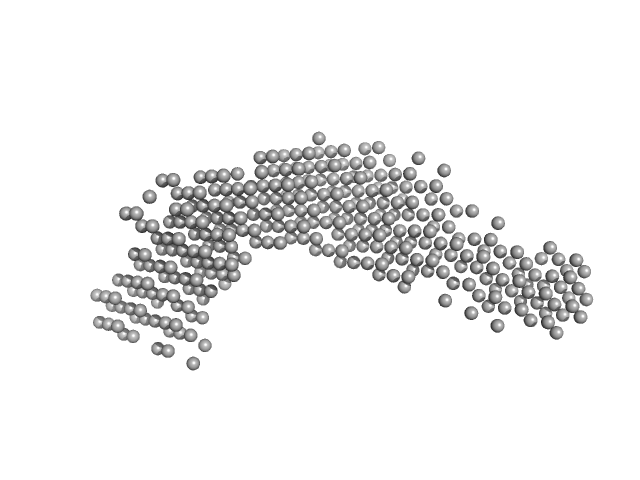
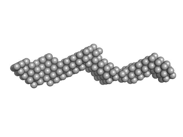
UniProt ID: Q15149-2 (543-1006) Plakin domain fragment of Human plectin encompassing spectrin repeats SR3-SR4-SR5-SR6 and SH3
|
|
|
|
| Sample: |
Plakin domain fragment of Human plectin encompassing spectrin repeats SR3-SR4-SR5-SR6 and SH3 monomer, 53 kDa Homo sapiens protein
|
| Buffer: |
20 mM Sodium Phosphate 150 mM NaCl 5% glycerol 3 mM DTT, pH: 7.5 |
| Experiment: |
SAXS
data collected at EMBL P12, PETRA III on 2013 Nov 26
|
The Structure of the Plakin Domain of Plectin Reveals an Extended Rod-like Shape.
J Biol Chem 291(36):18643-62 (2016)
Ortega E, Manso JA, Buey RM, Carballido AM, Carabias A, Sonnenberg A, de Pereda JM
|
| RgGuinier |
5.1 |
nm |
| Dmax |
21.0 |
nm |
| VolumePorod |
91 |
nm3 |
|
|
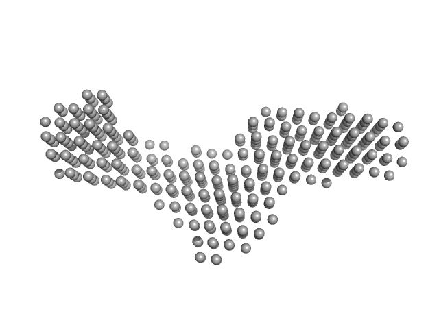
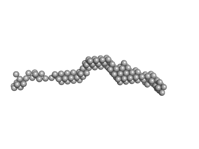
UniProt ID: Q15149-2 (1004-1372) Plakin domain fragment of Human plectin encompassing spectrin repeats SR7-SR8-SR9
|
|
|
|
| Sample: |
Plakin domain fragment of Human plectin encompassing spectrin repeats SR7-SR8-SR9 monomer, 43 kDa Homo sapiens protein
|
| Buffer: |
20 mM Sodium Phosphate 150 mM NaCl 5% glycerol 3 mM DTT, pH: 7.5 |
| Experiment: |
SAXS
data collected at EMBL P12, PETRA III on 2013 Nov 26
|
The Structure of the Plakin Domain of Plectin Reveals an Extended Rod-like Shape.
J Biol Chem 291(36):18643-62 (2016)
Ortega E, Manso JA, Buey RM, Carballido AM, Carabias A, Sonnenberg A, de Pereda JM
|
| RgGuinier |
4.2 |
nm |
| Dmax |
17.0 |
nm |
| VolumePorod |
57 |
nm3 |
|
|
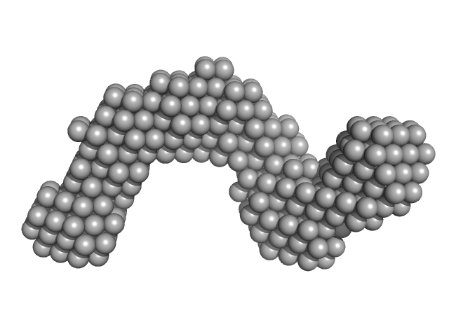
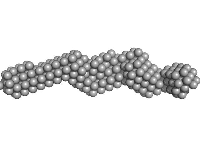
UniProt ID: Q15149-2 (543-1372) Plakin domain fragment of Human plectin encompassing spectrin repeats SR3-SR9
|
|
|
|
| Sample: |
Plakin domain fragment of Human plectin encompassing spectrin repeats SR3-SR9 monomer, 96 kDa Homo sapiens protein
|
| Buffer: |
20 mM Sodium Phosphate 150 mM NaCl 5% glycerol 3 mM DTT, pH: 7.5 |
| Experiment: |
SAXS
data collected at EMBL P12, PETRA III on 2013 Aug 13
|
The Structure of the Plakin Domain of Plectin Reveals an Extended Rod-like Shape.
J Biol Chem 291(36):18643-62 (2016)
Ortega E, Manso JA, Buey RM, Carballido AM, Carabias A, Sonnenberg A, de Pereda JM
|
| RgGuinier |
8.5 |
nm |
| Dmax |
35.0 |
nm |
| VolumePorod |
135 |
nm3 |
|
|
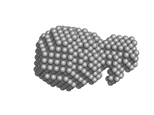
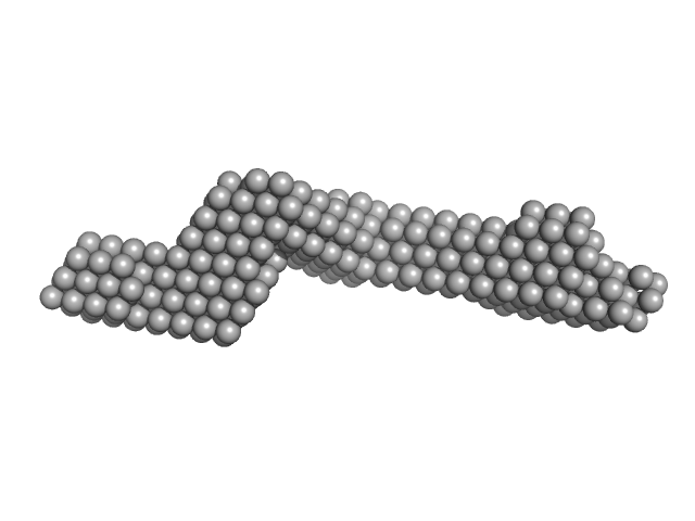
UniProt ID: P15924 (660-1025) Plakin domain fragment of Human Desmoplakin encompassing spectrin repeats SR7-SR8-SR9
|
|
|
|
| Sample: |
Plakin domain fragment of Human Desmoplakin encompassing spectrin repeats SR7-SR8-SR9 monomer, 42 kDa Homo sapiens protein
|
| Buffer: |
20 mM Sodium Phosphate 150 mM NaCl 5% glycerol 3 mM DTT, pH: 7.5 |
| Experiment: |
SAXS
data collected at EMBL P12, PETRA III on 2015 Sep 25
|
The Structure of the Plakin Domain of Plectin Reveals an Extended Rod-like Shape.
J Biol Chem 291(36):18643-62 (2016)
Ortega E, Manso JA, Buey RM, Carballido AM, Carabias A, Sonnenberg A, de Pereda JM
|
| RgGuinier |
4.4 |
nm |
| Dmax |
17.5 |
nm |
| VolumePorod |
69 |
nm3 |
|
|

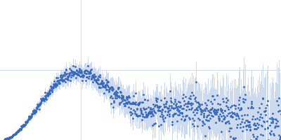

 experimental SAS data")


 from Proteus mirabilis Rg histogram")


