UniProt ID: Q6CUZ3 (1-481) Glucokinase-1
UniProt ID: Q6CUZ3 (1-481) Glucokinase-1
|
|
|
|
| Sample: |
Glucokinase-1 monomer, 54 kDa Kluyveromyces lactis (strain … protein
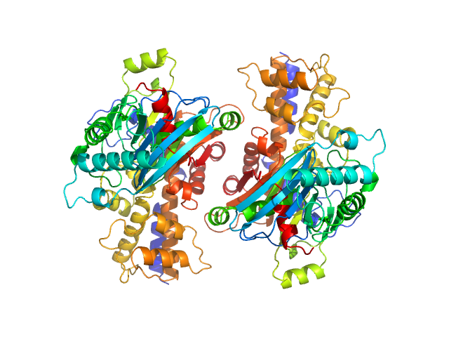
Glucokinase-1 dimer, 108 kDa Kluyveromyces lactis (strain … protein
|
| Buffer: |
10 mM Tris-HCl, 1 mM DTT, 10 mM D-fructose, pH: 7.4 |
| Experiment: |
SAXS
data collected at EMBL P12, PETRA III on 2017 Oct 13
|
Sugar binding to Kluyveromyces lactis glucokinase 1 (KlGlk1)
Renato H. Weiße
|
|
|
UniProt ID: Q6CUZ3 (1-481) Glucokinase-1
UniProt ID: Q6CUZ3 (1-481) Glucokinase-1
|
|
|

|
| Sample: |
Glucokinase-1 monomer, 54 kDa Kluyveromyces lactis (strain … protein
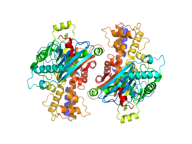
Glucokinase-1 dimer, 108 kDa Kluyveromyces lactis (strain … protein
|
| Buffer: |
10 mM Tris-HCl, 1 mM DTT, 10 mM D-mannose, pH: 7.4 |
| Experiment: |
SAXS
data collected at EMBL P12, PETRA III on 2017 Oct 13
|
Sugar binding to Kluyveromyces lactis glucokinase 1 (KlGlk1)
Renato H. Weiße
|
| RgGuinier |
2.7 |
nm |
| Dmax |
10.0 |
nm |
|
|
UniProt ID: Q6CUZ3 (1-481) Glucokinase-1 (H304Q)
UniProt ID: Q6CUZ3 (1-481) Glucokinase-1 (H304Q)
|
|
|
|
| Sample: |
Glucokinase-1 (H304Q) monomer, 54 kDa Kluyveromyces lactis (strain … protein
Glucokinase-1 (H304Q) dimer, 107 kDa Kluyveromyces lactis (strain … protein
|
| Buffer: |
10 mM Tris-HCl, 1 mM DTT, pH: 7.4 |
| Experiment: |
SAXS
data collected at EMBL P12, PETRA III on 2017 Oct 13
|
Sugar binding to Kluyveromyces lactis glucokinase 1 (KlGlk1)
Renato H. Weiße
|
|
|
UniProt ID: Q6CUZ3 (1-481) Glucokinase-1 (H304Q)
UniProt ID: Q6CUZ3 (1-481) Glucokinase-1 (H304Q)
|
|
|

|
| Sample: |
Glucokinase-1 (H304Q) monomer, 54 kDa Kluyveromyces lactis (strain … protein
Glucokinase-1 (H304Q) dimer, 107 kDa Kluyveromyces lactis (strain … protein
|
| Buffer: |
10 mM Tris-HCl, 1 mM DTT, 10 mM D-fructose, pH: 7.4 |
| Experiment: |
SAXS
data collected at EMBL P12, PETRA III on 2017 Oct 13
|
Sugar binding to Kluyveromyces lactis glucokinase 1 (KlGlk1)
Renato H. Weiße
|
|
|
UniProt ID: Q6CUZ3 (1-481) Glucokinase-1 (H304Q)
UniProt ID: Q6CUZ3 (1-481) Glucokinase-1 (H304Q)
|
|
|

|
| Sample: |
Glucokinase-1 (H304Q) monomer, 54 kDa Kluyveromyces lactis (strain … protein
Glucokinase-1 (H304Q) dimer, 107 kDa Kluyveromyces lactis (strain … protein
|
| Buffer: |
10 mM Tris-HCl, 1 mM DTT, 10 mM D-mannose, pH: 7.4 |
| Experiment: |
SAXS
data collected at EMBL P12, PETRA III on 2017 Oct 13
|
Sugar binding to Kluyveromyces lactis glucokinase 1 (KlGlk1)
Renato H. Weiße
|
|
|
UniProt ID: Q6CUZ3 (1-481) Glucokinase-1 (I356D, Y419D, H420D)
|
|
|
|
| Sample: |
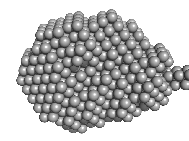
Glucokinase-1 (I356D, Y419D, H420D) monomer, 54 kDa Kluyveromyces lactis (strain … protein
|
| Buffer: |
10 mM Tris-HCl, 1 mM DTT, pH: 7.4 |
| Experiment: |
SAXS
data collected at EMBL P12, PETRA III on 2017 Oct 13
|
Sugar binding to Kluyveromyces lactis glucokinase 1 (KlGlk1)
Renato H. Weiße
|
| RgGuinier |
2.6 |
nm |
| Dmax |
9.0 |
nm |
| VolumePorod |
78 |
nm3 |
|
|
UniProt ID: Q6CUZ3 (1-481) Glucokinase-1 (I356D, Y419D, H420D)
|
|
|

|
| Sample: |
Glucokinase-1 (I356D, Y419D, H420D) monomer, 54 kDa Kluyveromyces lactis (strain … protein
|
| Buffer: |
10 mM Tris-HCl, 1 mM DTT, pH: 7.4 |
| Experiment: |
SAXS
data collected at EMBL P12, PETRA III on 2017 Oct 13
|
Sugar binding to Kluyveromyces lactis glucokinase 1 (KlGlk1)
Renato H. Weiße
|
| RgGuinier |
2.4 |
nm |
| Dmax |
8.0 |
nm |
| VolumePorod |
76 |
nm3 |
|
|
UniProt ID: Q6CUZ3 (1-481) Glucokinase-1 (I356D, Y419D, H420D)
|
|
|
|
| Sample: |
Glucokinase-1 (I356D, Y419D, H420D) monomer, 54 kDa Kluyveromyces lactis (strain … protein
|
| Buffer: |
10 mM Tris-HCl, 1 mM DTT, pH: 7.4 |
| Experiment: |
SAXS
data collected at EMBL P12, PETRA III on 2017 Oct 13
|
Sugar binding to Kluyveromyces lactis glucokinase 1 (KlGlk1)
Renato H. Weiße
|
| RgGuinier |
2.5 |
nm |
| Dmax |
8.0 |
nm |
| VolumePorod |
75 |
nm3 |
|
|
UniProt ID: Q6CUZ3 (1-481) Glucokinase-1 (I356D, Y419D, H420D)
|
|
|

|
| Sample: |
Glucokinase-1 (I356D, Y419D, H420D) monomer, 54 kDa Kluyveromyces lactis (strain … protein
|
| Buffer: |
10 mM Tris-HCl, 1 mM DTT, pH: 7.4 |
| Experiment: |
SAXS
data collected at EMBL P12, PETRA III on 2017 Oct 13
|
Sugar binding to Kluyveromyces lactis glucokinase 1 (KlGlk1)
Renato H. Weiße
|
|
|
UniProt ID: Q6CUZ3 (1-481) Glucokinase-1 (H304Q)
|
|
|

|
| Sample: |
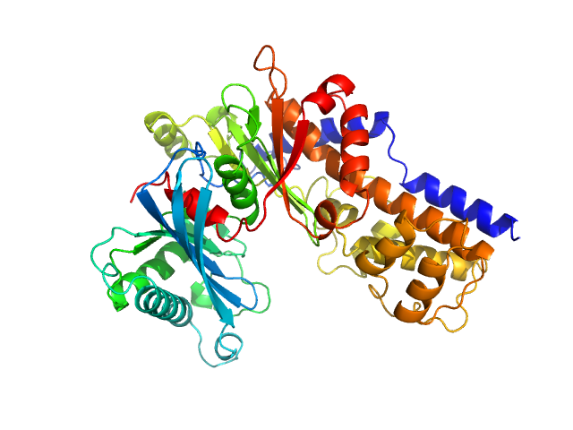
Glucokinase-1 (H304Q) monomer, 54 kDa Kluyveromyces lactis (strain … protein
|
| Buffer: |
10 mM Tris-HCl, 1 mM DTT, 10 mM D-fructose, pH: 7.4 |
| Experiment: |
SAXS
data collected at EMBL P12, PETRA III on 2018 May 14
|
Sugar binding to Kluyveromyces lactis glucokinase 1 (KlGlk1)
Renato H. Weiße
|
| RgGuinier |
2.5 |
nm |
| Dmax |
8.2 |
nm |
| VolumePorod |
76 |
nm3 |
|
|


Glucokinase-1 (H304Q) experimental SAS data")
Glucokinase-1 (H304Q) experimental SAS data")
Glucokinase-1 (H304Q) experimental SAS data")
 experimental SAS data")
 experimental SAS data")
 experimental SAS data")
 experimental SAS data")
 experimental SAS data")