UniProt ID: Q5ZTK4 (1158-1499) Ubiquitinating/deubiquitinating enzyme SdeA
|
|
|

|
| Sample: |
Ubiquitinating/deubiquitinating enzyme SdeA monomer, 39 kDa Legionella pneumophila subsp. … protein
|
| Buffer: |
25 mM Tris-HCl, 200 mM NaCl, 5 mM DTT, pH: 7.5 |
| Experiment: |
SAXS
data collected at EMBL P12, PETRA III on 2020 Dec 4
|
Structural Basis for Membrane Targeting and Secretion of Legionella SidE Ubiquitin Ligases
Ahmed Mohammed
|
| RgGuinier |
3.9 |
nm |
| Dmax |
15.7 |
nm |
| VolumePorod |
62 |
nm3 |
|
|
UniProt ID: Q5ZYD0 (1-111) Type 4 adapter protein IcmS
UniProt ID: Q5ZS31 (1-151) Type 4 adapter protein IcmW (S9A)
UniProt ID: Q5ZYC6 (656-783) Type 4 coupling protein DotL
|
|
|

|
| Sample: |
Type 4 adapter protein IcmS monomer, 14 kDa Legionella pneumophila subsp. … protein
Type 4 adapter protein IcmW (S9A) monomer, 17 kDa Legionella pneumophila subsp. … protein
Type 4 coupling protein DotL monomer, 15 kDa Legionella pneumophila subsp. … protein
|
| Buffer: |
25 mM Tris-HCl, 200 mM NaCl, 5 mM DTT, pH: 7.5 |
| Experiment: |
SAXS
data collected at EMBL P12, PETRA III on 2020 Dec 4
|
Structural Basis for Membrane Targeting and Secretion of Legionella SidE Ubiquitin Ligases
Ahmed Mohammed
|
| RgGuinier |
2.8 |
nm |
| Dmax |
9.6 |
nm |
| VolumePorod |
71 |
nm3 |
|
|
UniProt ID: Q5ZTK4 (1158-1499) Ubiquitinating/deubiquitinating enzyme SdeA
UniProt ID: Q5ZYD0 (1-111) Type 4 adapter protein IcmS
UniProt ID: Q5ZS31 (1-151) Type 4 adapter protein IcmW (S9A)
UniProt ID: Q5ZYC6 (656-783) Type 4 coupling protein DotL
|
|
|

|
| Sample: |
Ubiquitinating/deubiquitinating enzyme SdeA monomer, 39 kDa Legionella pneumophila subsp. … protein
Type 4 adapter protein IcmS monomer, 14 kDa Legionella pneumophila subsp. … protein
Type 4 adapter protein IcmW (S9A) monomer, 17 kDa Legionella pneumophila subsp. … protein
Type 4 coupling protein DotL monomer, 15 kDa Legionella pneumophila subsp. … protein
|
| Buffer: |
25 mM Tris-HCl, 200 mM NaCl, 5 mM DTT, pH: 7.5 |
| Experiment: |
SAXS
data collected at EMBL P12, PETRA III on 2020 Dec 4
|
Structural Basis for Membrane Targeting and Secretion of Legionella SidE Ubiquitin Ligases
Ahmed Mohammed
|
| RgGuinier |
3.8 |
nm |
| Dmax |
14.4 |
nm |
| VolumePorod |
166 |
nm3 |
|
|
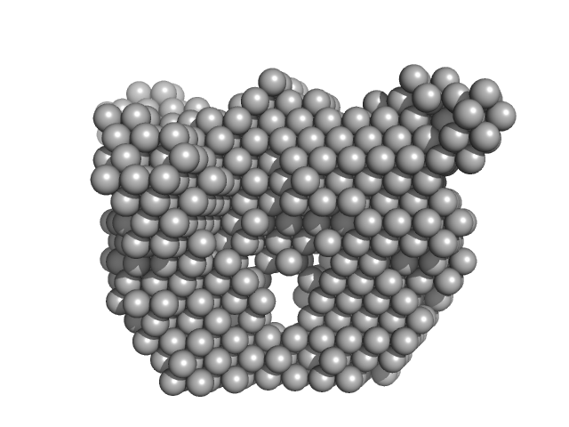
UniProt ID: Q6CUZ3 (1-481) Glucokinase-1
|
|
|
|
| Sample: |
Glucokinase-1 dimer, 108 kDa Kluyveromyces lactis (strain … protein
|
| Buffer: |
10 mM Tris-HCl, 1 mM DTT, pH: 7.4 |
| Experiment: |
SAXS
data collected at EMBL P12, PETRA III on 2015 Oct 20
|
Sugar binding to Kluyveromyces lactis glucokinase 1 (KlGlk1)
Renato H. Weiße
|
| RgGuinier |
3.4 |
nm |
| Dmax |
9.9 |
nm |
| VolumePorod |
154 |
nm3 |
|
|
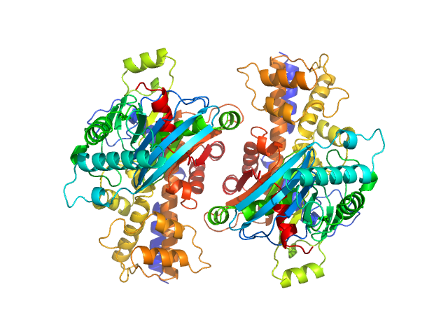
UniProt ID: Q6CUZ3 (1-481) Glucokinase-1
UniProt ID: Q6CUZ3 (1-481) Glucokinase-1
|
|
|
|
| Sample: |
Glucokinase-1 monomer, 54 kDa Kluyveromyces lactis (strain … protein
Glucokinase-1 dimer, 108 kDa Kluyveromyces lactis (strain … protein
|
| Buffer: |
10 mM Tris-HCl, 1 mM DTT, pH: 7.4 |
| Experiment: |
SAXS
data collected at EMBL P12, PETRA III on 2015 Oct 20
|
Sugar binding to Kluyveromyces lactis glucokinase 1 (KlGlk1)
Renato H. Weiße
|
| RgGuinier |
3.1 |
nm |
| Dmax |
9.5 |
nm |
| VolumePorod |
108 |
nm3 |
|
|
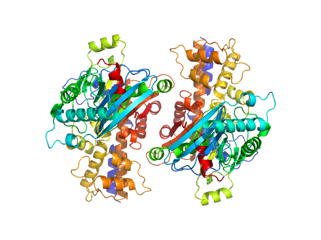
UniProt ID: Q6CUZ3 (1-481) Glucokinase-1
UniProt ID: Q6CUZ3 (1-481) Glucokinase-1
|
|
|
|
| Sample: |
Glucokinase-1 monomer, 54 kDa Kluyveromyces lactis (strain … protein
Glucokinase-1 dimer, 108 kDa Kluyveromyces lactis (strain … protein
|
| Buffer: |
10 mM Tris-HCl, 1 mM DTT, pH: 7.4 |
| Experiment: |
SAXS
data collected at EMBL P12, PETRA III on 2015 Oct 20
|
Sugar binding to Kluyveromyces lactis glucokinase 1 (KlGlk1)
Renato H. Weiße
|
| RgGuinier |
3.1 |
nm |
| Dmax |
9.5 |
nm |
| VolumePorod |
128 |
nm3 |
|
|
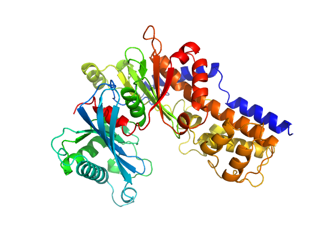
UniProt ID: Q6CUZ3 (1-481) Glucokinase-1
UniProt ID: Q6CUZ3 (1-481) Glucokinase-1
|
|
|

|
| Sample: |
Glucokinase-1 monomer, 54 kDa Kluyveromyces lactis (strain … protein
Glucokinase-1 dimer, 108 kDa Kluyveromyces lactis (strain … protein
|
| Buffer: |
10 mM Tris-HCl, 1 mM DTT, pH: 7.4 |
| Experiment: |
SAXS
data collected at EMBL P12, PETRA III on 2015 Oct 20
|
Sugar binding to Kluyveromyces lactis glucokinase 1 (KlGlk1)
Renato H. Weiße
|
| RgGuinier |
3.1 |
nm |
| Dmax |
10.0 |
nm |
| VolumePorod |
138 |
nm3 |
|
|
UniProt ID: Q6CUZ3 (1-481) Glucokinase-1
UniProt ID: Q6CUZ3 (1-481) Glucokinase-1
|
|
|

|
| Sample: |
Glucokinase-1 monomer, 54 kDa Kluyveromyces lactis (strain … protein
Glucokinase-1 dimer, 108 kDa Kluyveromyces lactis (strain … protein
|
| Buffer: |
10 mM Tris-HCl, 1 mM DTT, pH: 7.4 |
| Experiment: |
SAXS
data collected at EMBL P12, PETRA III on 2015 Oct 20
|
Sugar binding to Kluyveromyces lactis glucokinase 1 (KlGlk1)
Renato H. Weiße
|
| RgGuinier |
3.1 |
nm |
| Dmax |
10.0 |
nm |
| VolumePorod |
140 |
nm3 |
|
|
UniProt ID: Q6CUZ3 (1-481) Glucokinase-1
UniProt ID: Q6CUZ3 (1-481) Glucokinase-1
|
|
|
|
| Sample: |
Glucokinase-1 monomer, 54 kDa Kluyveromyces lactis (strain … protein
Glucokinase-1 dimer, 108 kDa Kluyveromyces lactis (strain … protein
|
| Buffer: |
10 mM Tris-HCl, 1 mM DTT, pH: 7.4 |
| Experiment: |
SAXS
data collected at EMBL P12, PETRA III on 2015 Oct 20
|
Sugar binding to Kluyveromyces lactis glucokinase 1 (KlGlk1)
Renato H. Weiße
|
| RgGuinier |
3.0 |
nm |
| Dmax |
10.0 |
nm |
|
|
UniProt ID: Q6CUZ3 (1-481) Glucokinase-1
UniProt ID: Q6CUZ3 (1-481) Glucokinase-1
|
|
|

|
| Sample: |
Glucokinase-1 monomer, 54 kDa Kluyveromyces lactis (strain … protein
Glucokinase-1 dimer, 108 kDa Kluyveromyces lactis (strain … protein
|
| Buffer: |
10 mM Tris-HCl, 1 mM DTT, pH: 7.4 |
| Experiment: |
SAXS
data collected at EMBL P12, PETRA III on 2015 Oct 20
|
Sugar binding to Kluyveromyces lactis glucokinase 1 (KlGlk1)
Renato H. Weiße
|
|
|

Type 4 coupling protein DotL experimental SAS data")
Type 4 coupling protein DotL experimental SAS data")


