|
|
|
|
|
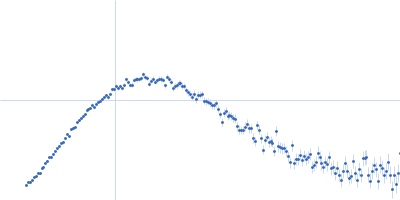
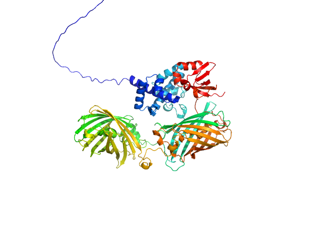
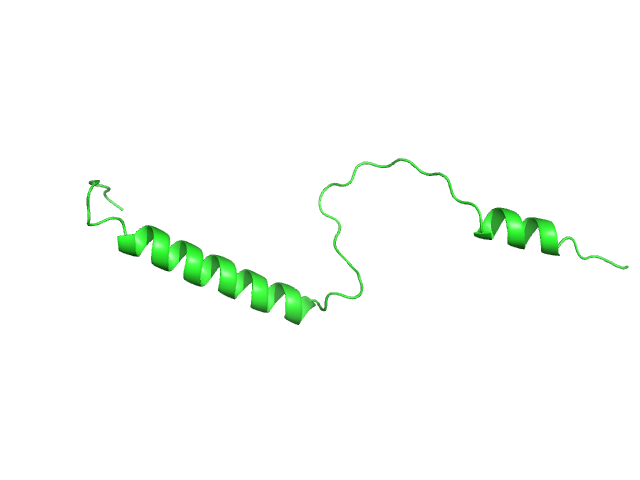
| Sample: |
RNA-directed RNA polymerase nsP4 monomer, 68 kDa O'nyong-nyong virus protein
|
| Buffer: |
25 mM HEPES, 1 mM TCEP, 5% glycerol and 500 mM NaCl., pH: 7.5 |
| Experiment: |
SAXS
data collected at Rigaku BioSAXS-2000, Pennsylvania State University on 2023 Jun 8
|
A fold switch regulates conformation of an alphavirus RNA-dependent RNA polymerase.
Nucleic Acids Res 54(2) (2026)
Arnold JJ, Braet SM, Vieira LC, Moustafa IM, Gohara DW, Fecko JA, Su YN, Jain A, Aponte-Diaz D, Wilke CO, Anand GS, Yennawar NH, Cameron CE
|
|
|
|
|
|
|
|
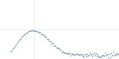
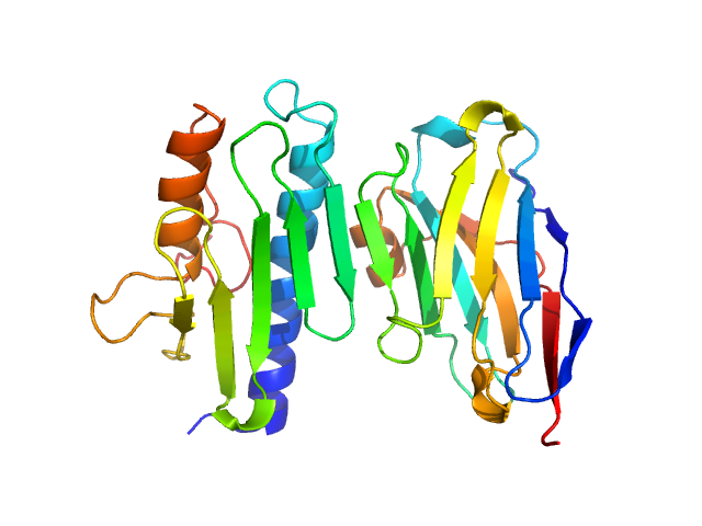
| Sample: |
50-residue-C-terminus-nsP3 fused with RNA-directed RNA polymerase nsP4 tetramer, 318 kDa O'nyong nyong virus protein
|
| Buffer: |
25 mM HEPES 5% glycerol, 1 mM TCEP, 500 mM NaCl., pH: 7.5 |
| Experiment: |
SAXS
data collected at Rigaku BioSAXS-2000, Pennsylvania State University on 2025 Aug 14
|
A fold switch regulates conformation of an alphavirus RNA-dependent RNA polymerase.
Nucleic Acids Res 54(2) (2026)
Arnold JJ, Braet SM, Vieira LC, Moustafa IM, Gohara DW, Fecko JA, Su YN, Jain A, Aponte-Diaz D, Wilke CO, Anand GS, Yennawar NH, Cameron CE
|
|
|
|
|
|
|
|
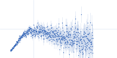
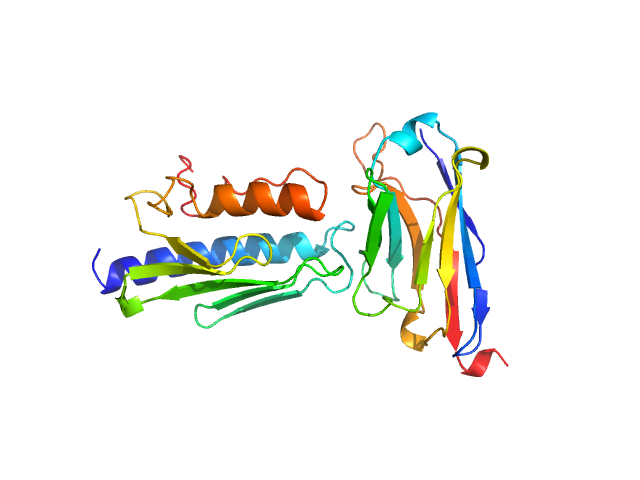
| Sample: |
Ratiometric matryoshka biosensor for Fe2+ monomer, 84 kDa synthetic construct protein
|
| Buffer: |
20 mM MOPS, 250 mM KCl,, pH: 7 |
| Experiment: |
SAXS
data collected at BM29, ESRF on 2024 Nov 15
|
A Novel Biosensor for Ferrous Iron Developed via CoBiSe: A Computational Method for Rapid Biosensor Design
ACS Sensors (2026)
Papadopoulos A, Anlauf M, Reiners J, Paik S, Krüger A, Lückel B, Bott M, Drepper T, Frunzke J, Gohlke H, Weidtkamp-Peters S, Smits S, Gertzen C
|
| RgGuinier |
3.6 |
nm |
| Dmax |
12.4 |
nm |
| VolumePorod |
118 |
nm3 |
|
|
|
|
|
|

|
| Sample: |
Isoform Short of Small EDRK-rich factor 1 monomer, 7 kDa Homo sapiens protein
|
| Buffer: |
20 mM sodium phosphate, 20 mM NaCl, pH: 6 |
| Experiment: |
SAXS
data collected at 13A, Taiwan Photon Source, NSRRC on 2023 Mar 9
|
pH Sensitivity of the SERF1a Conformational Ensemble
ACS Omega (2026)
Huang S, Shih O, Jeng U, Chang C, Lin J, Malliavin T
|
| RgGuinier |
2.4 |
nm |
| Dmax |
10.3 |
nm |
| VolumePorod |
10 |
nm3 |
|
|
|
|
|
|
|
| Sample: |
Isoform Short of Small EDRK-rich factor 1 monomer, 7 kDa Homo sapiens protein
|
| Buffer: |
20 mM sodium phosphate, 20 mM NaCl, pH: 6.8 |
| Experiment: |
SAXS
data collected at 13A, Taiwan Photon Source, NSRRC on 2023 Mar 9
|
pH Sensitivity of the SERF1a Conformational Ensemble
ACS Omega (2026)
Huang S, Shih O, Jeng U, Chang C, Lin J, Malliavin T
|
| RgGuinier |
2.5 |
nm |
| Dmax |
8.8 |
nm |
| VolumePorod |
11 |
nm3 |
|
|
|
|
|
|
|
| Sample: |
Frataxin, mitochondrial monomer, 14 kDa Homo sapiens protein
Nanobody 28F6 monomer, 15 kDa synthetic construct protein
|
| Buffer: |
20 mM Tris, 150 mM NaCl, pH: 7.5 |
| Experiment: |
SAXS
data collected at B21, Diamond Light Source on 2024 Sep 18
|
Nanobodies as tools for studying human frataxin biology.
Commun Biol (2026)
Pignataro MF, Fernández NB, Garay-Alvarez A, Pavan MF, Molina R, Muñoz IG, Grossi J, Noguera M, Vila A, García AE, Gentili HG, Rodríguez NA, Aran M, Parreño V, Bok M, Hermoso JA, Ibañez LI, Santos J
|
| RgGuinier |
2.2 |
nm |
| Dmax |
7.4 |
nm |
| VolumePorod |
36 |
nm3 |
|
|
|
|
|
|
|
| Sample: |
Frataxin, mitochondrial monomer, 14 kDa Homo sapiens protein
Nanobody 16C10 monomer, 15 kDa synthetic construct protein
|
| Buffer: |
20 mM Tris, 150 mM NaCl, pH: 7.5 |
| Experiment: |
SAXS
data collected at B21, Diamond Light Source on 2024 Sep 18
|
Nanobodies as tools for studying human frataxin biology.
Commun Biol (2026)
Pignataro MF, Fernández NB, Garay-Alvarez A, Pavan MF, Molina R, Muñoz IG, Grossi J, Noguera M, Vila A, García AE, Gentili HG, Rodríguez NA, Aran M, Parreño V, Bok M, Hermoso JA, Ibañez LI, Santos J
|
| RgGuinier |
2.4 |
nm |
| Dmax |
8.0 |
nm |
| VolumePorod |
37 |
nm3 |
|
|
|
|
|
|
|
| Sample: |
Pigeon iron-sulfur cluster assembly 1 homolog, mitochondrial, 15 kDa Columba livia protein
|
| Buffer: |
20 mM Tris-HCl, 0.15 M NaCl, 10 mM 3-mercapto-1,2-propanediol, pH: 8 |
| Experiment: |
SAXS
data collected at BL-10C, Photon Factory (PF), High Energy Accelerator Research Organization (KEK) on 2021 Jun 8
|
A hidden property of the iron-sulfur protein in the mononuclear iron-bound state: species-dependent structural ordering induced by magnetic fields.
FEBS J (2025)
Arai S, Soga S, Hirai M, Kobayashi R, Masai H, Kimura K, Maeda K, Nagashima H
|
| RgGuinier |
2.8 |
nm |
| Dmax |
10.5 |
nm |
| VolumePorod |
85 |
nm3 |
|
|
|
|
|
|
|
| Sample: |
Human Iron-sulfur cluster assembly 1 homolog, mitochondrial, 14 kDa Homo sapiens protein
|
| Buffer: |
20 mM Tris-HCl, 0.15 M NaCl, 10 mM 3-mercapto-1,2-propanediol, pH: 8 |
| Experiment: |
SAXS
data collected at BL-10C, Photon Factory (PF), High Energy Accelerator Research Organization (KEK) on 2024 Apr 29
|
A hidden property of the iron-sulfur protein in the mononuclear iron-bound state: species-dependent structural ordering induced by magnetic fields.
FEBS J (2025)
Arai S, Soga S, Hirai M, Kobayashi R, Masai H, Kimura K, Maeda K, Nagashima H
|
| RgGuinier |
4.1 |
nm |
| Dmax |
16.4 |
nm |
| VolumePorod |
105 |
nm3 |
|
|
|
|
|
|
|
| Sample: |
Human Iron-sulfur cluster assembly 1 homolog, mitochondrial, 14 kDa Homo sapiens protein
|
| Buffer: |
20 mM Tris-HCl, 0.15 M NaCl, 10 mM 3-mercapto-1,2-propanediol, pH: 8 |
| Experiment: |
SAXS
data collected at BL-10C, Photon Factory (PF), High Energy Accelerator Research Organization (KEK) on 2023 Dec 1
|
A hidden property of the iron-sulfur protein in the mononuclear iron-bound state: species-dependent structural ordering induced by magnetic fields.
FEBS J (2025)
Arai S, Soga S, Hirai M, Kobayashi R, Masai H, Kimura K, Maeda K, Nagashima H
|
| RgGuinier |
2.9 |
nm |
| Dmax |
8.8 |
nm |
| VolumePorod |
55 |
nm3 |
|
|