|
|
|
|
|
| Sample: |
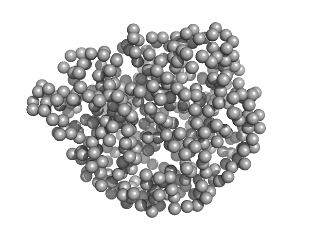
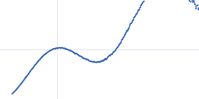
Lanthionine synthetase C-like protein monomer, 50 kDa Clostridium sp. Maddingley … protein
|
| Buffer: |
50 mM HEPES, 500 mM NaCl, pH: 8 |
| Experiment: |
SAXS
data collected at Xenocs Xeuss 2.0 Q-Xoom, Center for Structural Studies, Heinrich-Heine-University on 2019 Nov 28
|
The structure of MadC from Clostridium maddingley reveals new insights into class I lanthipeptide cyclases
Frontiers in Microbiology 13 (2023)
Knospe C, Kamel M, Spitz O, Hoeppner A, Galle S, Reiners J, Kedrov A, Smits S, Schmitt L
|
| RgGuinier |
2.3 |
nm |
| Dmax |
7.8 |
nm |
| VolumePorod |
78 |
nm3 |
|
|
|
|
|
|
|
| Sample: |
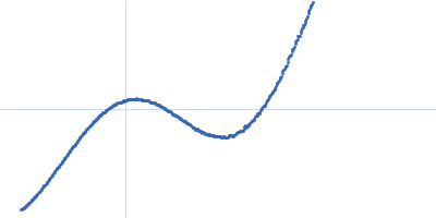
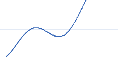
Alpha cyclodextrin monomer, 1 kDa
|
| Buffer: |
water, pH: 7 |
| Experiment: |
SAXS
data collected at 11-ID-B, Advanced Photon Source (APS), Argonne National Laboratory on 2021 Nov 2
|
Extended q‐range X‐ray Scattering Reveals High‐Resolution Structural Details of Biomacromolecules in Aqueous Solutions
Chemistry – A European Journal (2023)
Chen J, Borkiewicz O, Grishaev A, Zhang F, Bera M, Ruett U, Levin I
|
|
|
|
|
|
|
|
| Sample: |
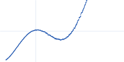
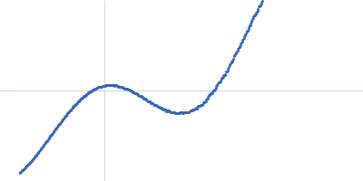
Alpha cyclodextrin monomer, 1 kDa
|
| Buffer: |
water, pH: 7 |
| Experiment: |
SAXS
data collected at 11-ID-B, Advanced Photon Source (APS), Argonne National Laboratory on 2021 Nov 2
|
Extended q‐range X‐ray Scattering Reveals High‐Resolution Structural Details of Biomacromolecules in Aqueous Solutions
Chemistry – A European Journal (2023)
Chen J, Borkiewicz O, Grishaev A, Zhang F, Bera M, Ruett U, Levin I
|
|
|
|
|
|
|
|
| Sample: |
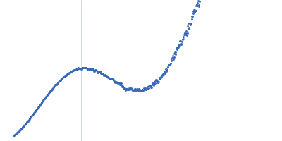
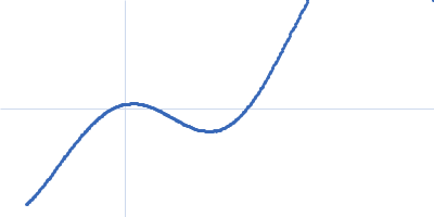
Alpha cyclodextrin monomer, 1 kDa
|
| Buffer: |
water, pH: 7 |
| Experiment: |
SAXS
data collected at 11-ID-B, Advanced Photon Source (APS), Argonne National Laboratory on 2021 Nov 2
|
Extended q‐range X‐ray Scattering Reveals High‐Resolution Structural Details of Biomacromolecules in Aqueous Solutions
Chemistry – A European Journal (2023)
Chen J, Borkiewicz O, Grishaev A, Zhang F, Bera M, Ruett U, Levin I
|
|
|
|
|
|
|
|
| Sample: |
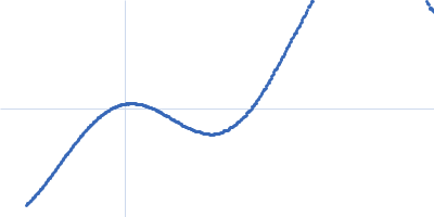
Beta Cyclodextrin monomer, 1 kDa
|
| Buffer: |
water, pH: 7 |
| Experiment: |
SAXS
data collected at 11-ID-B, Advanced Photon Source (APS), Argonne National Laboratory on 2021 Nov 2
|
Extended q‐range X‐ray Scattering Reveals High‐Resolution Structural Details of Biomacromolecules in Aqueous Solutions
Chemistry – A European Journal (2023)
Chen J, Borkiewicz O, Grishaev A, Zhang F, Bera M, Ruett U, Levin I
|
|
|
|
|
|
|
|
| Sample: |
Gamma Cyclodextrin monomer, 1 kDa
|
| Buffer: |
water, pH: 7 |
| Experiment: |
SAXS
data collected at 11-ID-B, Advanced Photon Source (APS), Argonne National Laboratory on 2021 Nov 2
|
Extended q‐range X‐ray Scattering Reveals High‐Resolution Structural Details of Biomacromolecules in Aqueous Solutions
Chemistry – A European Journal (2023)
Chen J, Borkiewicz O, Grishaev A, Zhang F, Bera M, Ruett U, Levin I
|
|
|
|
|
|
|
|
| Sample: |
His-tagged fused complex of cytochrome P450 143 and ferredoxin Rv1786 monomer, 54 kDa Mycobacterium tuberculosis protein
|
| Buffer: |
300 mM NaCl, 50 mM Tris/TrisHCl, 10% glycerol, pH: 7.4 |
| Experiment: |
SAXS
data collected at BM29, ESRF on 2018 Dec 7
|
Structural insights into 3Fe–4S ferredoxins diversity in M. tuberculosis highlighted by a first redox complex with P450
Frontiers in Molecular Biosciences 9 (2023)
Gilep A, Varaksa T, Bukhdruker S, Kavaleuski A, Ryzhykau Y, Smolskaya S, Sushko T, Tsumoto K, Grabovec I, Kapranov I, Okhrimenko I, Marin E, Shevtsov M, Mishin A, Kovalev K, Kuklin A, Gordeliy V, Kaluzhskiy L, Gnedenko O, Yablokov E, Ivanov A, Borshchevskiy V, Strushkevich N
|
| RgGuinier |
2.5 |
nm |
| Dmax |
9.0 |
nm |
| VolumePorod |
85 |
nm3 |
|
|


 Rg histogram")