|
|
|
|
|
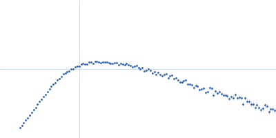
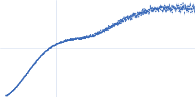
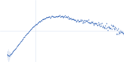
| Sample: |
Contactin-1 I433V dimer, 220 kDa Mus musculus protein
|
| Buffer: |
25 mM HEPES, 150 mM NaCl, pH: 7.5 |
| Experiment: |
SAXS
data collected at B21, Diamond Light Source on 2019 Dec 16
|
Structural insights into the contactin 1 – neurofascin 155 adhesion complex
Nature Communications 13(1) (2022)
Chataigner L, Gogou C, den Boer M, Frias C, Thies-Weesie D, Granneman J, Heck A, Meijer D, Janssen B
|
| RgGuinier |
8.0 |
nm |
| Dmax |
25.7 |
nm |
| VolumePorod |
350 |
nm3 |
|
|
|
|
|
|
|
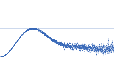
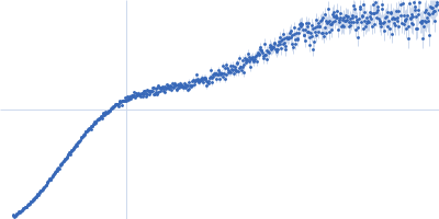
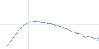
| Sample: |
Contactin-1 I433V dimer, 220 kDa Mus musculus protein
|
| Buffer: |
25 mM HEPES, 150 mM NaCl, pH: 7.5 |
| Experiment: |
SAXS
data collected at B21, Diamond Light Source on 2019 Dec 16
|
Structural insights into the contactin 1 – neurofascin 155 adhesion complex
Nature Communications 13(1) (2022)
Chataigner L, Gogou C, den Boer M, Frias C, Thies-Weesie D, Granneman J, Heck A, Meijer D, Janssen B
|
| RgGuinier |
7.0 |
nm |
| Dmax |
34.0 |
nm |
| VolumePorod |
285 |
nm3 |
|
|
|
|
|
|
|
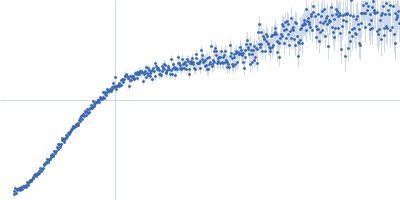
| Sample: |
Contactin-1 I433V dimer, 220 kDa Mus musculus protein
|
| Buffer: |
25 mM HEPES, 150 mM NaCl, pH: 7.5 |
| Experiment: |
SAXS
data collected at B21, Diamond Light Source on 2019 Dec 16
|
Structural insights into the contactin 1 – neurofascin 155 adhesion complex
Nature Communications 13(1) (2022)
Chataigner L, Gogou C, den Boer M, Frias C, Thies-Weesie D, Granneman J, Heck A, Meijer D, Janssen B
|
| RgGuinier |
6.8 |
nm |
| Dmax |
27.0 |
nm |
| VolumePorod |
275 |
nm3 |
|
|
|
|
|
|
|
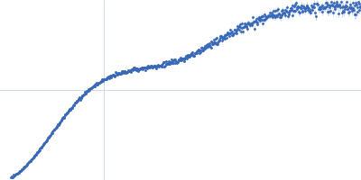
| Sample: |
Contactin-1 I433V dimer, 220 kDa Mus musculus protein
|
| Buffer: |
25 mM HEPES, 150 mM NaCl, pH: 7.5 |
| Experiment: |
SAXS
data collected at B21, Diamond Light Source on 2019 Dec 16
|
Structural insights into the contactin 1 – neurofascin 155 adhesion complex
Nature Communications 13(1) (2022)
Chataigner L, Gogou C, den Boer M, Frias C, Thies-Weesie D, Granneman J, Heck A, Meijer D, Janssen B
|
| RgGuinier |
6.8 |
nm |
| Dmax |
27.0 |
nm |
| VolumePorod |
270 |
nm3 |
|
|
|
|
|
|
|
| Sample: |
Metacaspase-1 monomer, 46 kDa Candida glabrata (strain … protein
|
| Buffer: |
10 mM HEPES, 150 mM NaCl, 1% glycerol, 10 mM CaCl2, pH: 7.6 |
| Experiment: |
SAXS
data collected at SWING, SOLEIL on 2019 Sep 26
|
Structural and molecular determinants of Candida glabrata metacaspase maturation and activation by calcium.
Commun Biol 5(1):1158 (2022)
Conchou L, Doumèche B, Galisson F, Violot S, Dugelay C, Diesis E, Page A, Bienvenu AL, Picot S, Aghajari N, Ballut L
|
| RgGuinier |
1.9 |
nm |
| Dmax |
5.4 |
nm |
| VolumePorod |
43 |
nm3 |
|
|
|
|
|
|
|
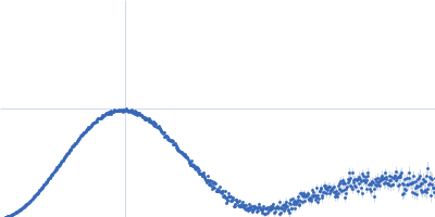
| Sample: |
Obscurin monomer, 21 kDa Homo sapiens protein
|
| Buffer: |
20 mM Tris, 50 mM NaCl, 0.35 mM NaN3, pH: 7.5 |
| Experiment: |
SAXS
data collected at 12.3.1 (SIBYLS), Advanced Light Source (ALS) on 2021 May 18
|
The N-terminus of obscurin is flexible in solution.
Proteins (2022)
Mauriello GE, Moncure GE, Nowzari RA, Miller CJ, Wright NT
|
| RgGuinier |
2.8 |
nm |
| Dmax |
10.5 |
nm |
| VolumePorod |
28 |
nm3 |
|
|
|
|
|
|
|
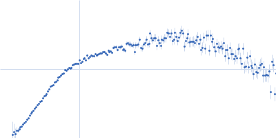
| Sample: |
Obscurin monomer, 21 kDa Homo sapiens protein
|
| Buffer: |
20 mM Tris, 50 mM NaCl, 0.35 mM NaN3, pH: 7.5 |
| Experiment: |
SAXS
data collected at 12.3.1 (SIBYLS), Advanced Light Source (ALS) on 2020 Nov 18
|
The N-terminus of obscurin is flexible in solution.
Proteins (2022)
Mauriello GE, Moncure GE, Nowzari RA, Miller CJ, Wright NT
|
| RgGuinier |
3.7 |
nm |
| Dmax |
18.0 |
nm |
| VolumePorod |
66 |
nm3 |
|
|
|
|
|
|
|
| Sample: |
Probable global transcription activator SNF2L2 (isoform 1) monomer, 16 kDa Homo sapiens protein
Von Hippel-Lindau disease tumor suppressor monomer, 19 kDa Homo sapiens protein
Elongin-B monomer, 12 kDa Homo sapiens protein
Elongin-C monomer, 11 kDa Homo sapiens protein
ACBI1 protac monomer, 1 kDa
|
| Buffer: |
20 mM HEPES, 150 mM NaCl, 1 mM DTT, pH: 7.5 |
| Experiment: |
SAXS
data collected at Xenocs BioXolver L with MetalJet, Département de Biochimie, Université de Montréal on 2021 Aug 11
|
Predicting the structural basis of targeted protein degradation by integrating molecular dynamics simulations with structural mass spectrometry.
Nat Commun 13(1):5884 (2022)
Dixon T, MacPherson D, Mostofian B, Dauzhenka T, Lotz S, McGee D, Shechter S, Shrestha UR, Wiewiora R, McDargh ZA, Pei F, Pal R, Ribeiro JV, Wilkerson T, Sachdeva V, Gao N, Jain S, Sparks S, Li Y, Vinitsky A, Zhang X, Razavi AM, Kolossváry I, Imbriglio J, Evdokimov A, Bergeron L, Zhou W, Adhikari J, Ruprecht B, Dickson A, Xu H, Sherman W, Izaguirre JA
|
| RgGuinier |
3.3 |
nm |
| Dmax |
12.5 |
nm |
| VolumePorod |
83 |
nm3 |
|
|
|
|
|
|
|
| Sample: |
Von Hippel-Lindau disease tumor suppressor monomer, 19 kDa Homo sapiens protein
Elongin-B monomer, 12 kDa Homo sapiens protein
Elongin-C monomer, 11 kDa Homo sapiens protein
ACBI1 protac monomer, 1 kDa
Probable global transcription activator SNF2L2 (isoform 2) monomer, 14 kDa Homo sapiens protein
|
| Buffer: |
20 mM HEPES, 150 mM NaCl, 1 mM DTT, pH: 7.5 |
| Experiment: |
SAXS
data collected at Xenocs BioXolver L with MetalJet, Département de Biochimie, Université de Montréal on 2021 Aug 11
|
Predicting the structural basis of targeted protein degradation by integrating molecular dynamics simulations with structural mass spectrometry.
Nat Commun 13(1):5884 (2022)
Dixon T, MacPherson D, Mostofian B, Dauzhenka T, Lotz S, McGee D, Shechter S, Shrestha UR, Wiewiora R, McDargh ZA, Pei F, Pal R, Ribeiro JV, Wilkerson T, Sachdeva V, Gao N, Jain S, Sparks S, Li Y, Vinitsky A, Zhang X, Razavi AM, Kolossváry I, Imbriglio J, Evdokimov A, Bergeron L, Zhou W, Adhikari J, Ruprecht B, Dickson A, Xu H, Sherman W, Izaguirre JA
|
| RgGuinier |
3.2 |
nm |
| Dmax |
11.1 |
nm |
| VolumePorod |
75 |
nm3 |
|
|
|
|
|
|
|
| Sample: |
Ribonuclease pancreatic monomer, 14 kDa Bos taurus protein
|
| Buffer: |
50 mM HEPES, 150 mM KCl, 3% glycerol, pH: 7.5 |
| Experiment: |
SAXS
data collected at EMBL P12, PETRA III on 2019 Sep 16
|
Small-Angle X-ray Scattering data (benchmarking/consensus): EMBL-P12 SAXS beam line, DESY
Clement Blanchet,
Melissa Graewert,
Cy M Jeffries,
Dmitri Svergun
|
| RgGuinier |
1.5 |
nm |
| Dmax |
4.6 |
nm |
| VolumePorod |
16 |
nm3 |
|
|


von Hippel-Lindau disease tumor suppressorElongin-BElongin-CACBI1 protac experimental SAS data")
 experimental SAS data")