|
|
|
|
|
| Sample: |
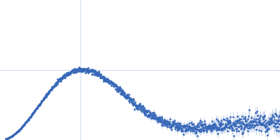
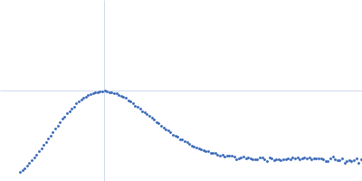
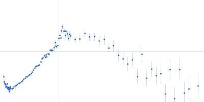
Phage-encoded SAM lyase Svi3-3 (including N-terminal His6-tag and Tev cleavage site) trimer, 56 kDa Unknown environmental phage protein
|
| Buffer: |
25 mM Tris-HCl, 150 mM NaCl, 5 mM SAM, pH: 8 |
| Experiment: |
SAXS
data collected at B21, Diamond Light Source on 2016 Dec 9
|
Structure and mechanism of a phage-encoded SAM lyase revises catalytic function of enzyme family.
Elife 10 (2021)
Guo X, Söderholm A, Kanchugal P S, Isaksen GV, Warsi O, Eckhard U, Trigüis S, Gogoll A, Jerlström-Hultqvist J, Åqvist J, Andersson DI, Selmer M
|
| RgGuinier |
2.5 |
nm |
| Dmax |
9.1 |
nm |
| VolumePorod |
89 |
nm3 |
|
|
|
|
|
|
|
| Sample: |
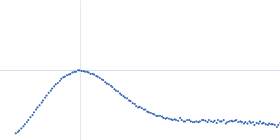
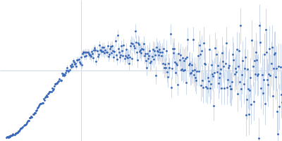
Apolipoprotein D tetramer, 77 kDa Homo sapiens protein
|
| Buffer: |
50 mM Na Phosphate, 150 mM NaCl, 3% glycerol, pH: 7.4 |
| Experiment: |
SAXS
data collected at SAXS/WAXS, Australian Synchrotron on 2016 Nov 11
|
Small angle X-ray scattering analysis of ligand-bound forms of tetrameric apolipoprotein-D
Bioscience Reports 41(1) (2021)
Kielkopf C, Whitten A, Garner B, Brown S
|
| RgGuinier |
3.3 |
nm |
| Dmax |
10.5 |
nm |
| VolumePorod |
168 |
nm3 |
|
|
|
|
|
|
|
| Sample: |
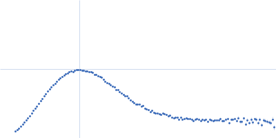
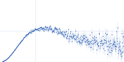
Apolipoprotein D tetramer, 77 kDa Homo sapiens protein
|
| Buffer: |
50 mM Na Phosphate, 150 mM NaCl, 3% glycerol, pH: 7.4 |
| Experiment: |
SAXS
data collected at SAXS/WAXS, Australian Synchrotron on 2016 Nov 11
|
Small angle X-ray scattering analysis of ligand-bound forms of tetrameric apolipoprotein-D
Bioscience Reports 41(1) (2021)
Kielkopf C, Whitten A, Garner B, Brown S
|
| RgGuinier |
3.3 |
nm |
| Dmax |
10.3 |
nm |
| VolumePorod |
164 |
nm3 |
|
|
|
|
|
|
|
| Sample: |
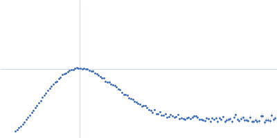
Apolipoprotein D tetramer, 77 kDa Homo sapiens protein
|
| Buffer: |
50 mM Na Phosphate, 150 mM NaCl, 3% glycerol, pH: 7.4 |
| Experiment: |
SAXS
data collected at SAXS/WAXS, Australian Synchrotron on 2016 Nov 11
|
Small angle X-ray scattering analysis of ligand-bound forms of tetrameric apolipoprotein-D
Bioscience Reports 41(1) (2021)
Kielkopf C, Whitten A, Garner B, Brown S
|
| RgGuinier |
3.3 |
nm |
| Dmax |
10.6 |
nm |
| VolumePorod |
163 |
nm3 |
|
|
|
|
|
|
|
| Sample: |
Apolipoprotein D tetramer, 77 kDa Homo sapiens protein
|
| Buffer: |
50 mM Na Phosphate, 150 mM NaCl, 3% glycerol, pH: 7.4 |
| Experiment: |
SAXS
data collected at SAXS/WAXS, Australian Synchrotron on 2016 Nov 11
|
Small angle X-ray scattering analysis of ligand-bound forms of tetrameric apolipoprotein-D
Bioscience Reports 41(1) (2021)
Kielkopf C, Whitten A, Garner B, Brown S
|
| RgGuinier |
3.3 |
nm |
| Dmax |
10.0 |
nm |
| VolumePorod |
161 |
nm3 |
|
|
|
|
|
|
|
| Sample: |
Apolipoprotein D tetramer, 77 kDa Homo sapiens protein
|
| Buffer: |
50 mM Na Phosphate, 150 mM NaCl, 3% glycerol, pH: 7.4 |
| Experiment: |
SAXS
data collected at SAXS/WAXS, Australian Synchrotron on 2016 Nov 11
|
Small angle X-ray scattering analysis of ligand-bound forms of tetrameric apolipoprotein-D
Bioscience Reports 41(1) (2021)
Kielkopf C, Whitten A, Garner B, Brown S
|
| RgGuinier |
3.3 |
nm |
| Dmax |
9.9 |
nm |
| VolumePorod |
171 |
nm3 |
|
|
|
|
|
|
|
| Sample: |
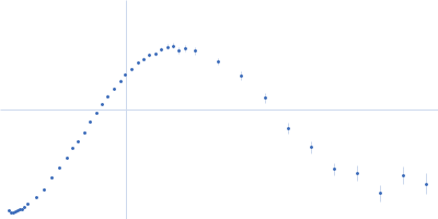
GATA-type iron responsive transcription factor Fep1 reconstituted monomer, 22 kDa Komagataella pastoris protein
24-mer double strand DNA from the GATA promoter dimer, 14 kDa synthetic oligonucleotide DNA
|
| Buffer: |
50 mM MOPS, 50 mM NaCl, pH: 7 |
| Experiment: |
SAXS
data collected at BM29, ESRF on 2018 Dec 5
|
Biophysical characterization of the complex between the iron-responsive transcription factor Fep1 and DNA.
Eur Biophys J (2021)
Miele AE, Cervoni L, Le Roy A, Cutone A, Musci G, Ebel C, Bonaccorsi di Patti MC
|
| RgGuinier |
3.5 |
nm |
| Dmax |
13.1 |
nm |
| VolumePorod |
73 |
nm3 |
|
|
|
|
|
|
|
| Sample: |
GATA-type iron responsive transcription factor Fep1 monomer, 22 kDa Komagataella pastoris protein
24-mer double strand DNA from the GATA promoter dimer, 14 kDa synthetic oligonucleotide DNA
|
| Buffer: |
50 mM MOPS, 50 mM NaCl, pH: 7 |
| Experiment: |
SAXS
data collected at BM29, ESRF on 2018 Dec 5
|
Biophysical characterization of the complex between the iron-responsive transcription factor Fep1 and DNA.
Eur Biophys J (2021)
Miele AE, Cervoni L, Le Roy A, Cutone A, Musci G, Ebel C, Bonaccorsi di Patti MC
|
| RgGuinier |
3.2 |
nm |
| Dmax |
12.0 |
nm |
| VolumePorod |
85 |
nm3 |
|
|
|
|
|
|
|
| Sample: |
Bacteriorhodopsin monomer, 27 kDa Halobacterium salinarum protein
|
| Buffer: |
25 mM NaH2PO4, 1.35 mM KOH, 40 mM partially-deuterated octyl glucoside mixture, pH: 5.6 |
| Experiment: |
SANS
data collected at NGB 30m SANS, NIST Center for High Resolution Neutron Scattering (CHRNS) on 2017 Jan 20
|
Direct localization of detergents and bacteriorhodopsin in the lipidic cubic phase by small-angle neutron scattering
IUCrJ 8(1) (2021)
Cleveland IV T, Blick E, Krueger S, Leung A, Darwish T, Butler P
|
| RgGuinier |
2.7 |
nm |
| Dmax |
5.4 |
nm |
| VolumePorod |
68 |
nm3 |
|
|
|
|
|
|
|
| Sample: |
Bacteriorhodopsin monomer, 27 kDa Halobacterium salinarum protein
|
| Buffer: |
25 mM NaH2PO4, 1.35 mM KOH, 40 mM octyl glucoside, pH: 5.6 |
| Experiment: |
SANS
data collected at NG7, NIST Center for High Resolution Neutron Scattering (CHRNS) on 2017 May 15
|
Direct localization of detergents and bacteriorhodopsin in the lipidic cubic phase by small-angle neutron scattering
IUCrJ 8(1) (2021)
Cleveland IV T, Blick E, Krueger S, Leung A, Darwish T, Butler P
|
| RgGuinier |
2.7 |
nm |
| Dmax |
6.2 |
nm |
| VolumePorod |
49 |
nm3 |
|
|
 experimental SAS data")