|
|
|
|
|
| Sample: |
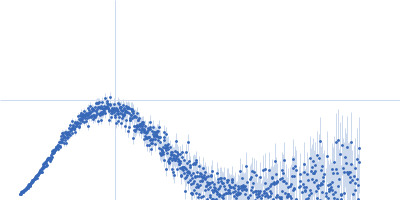
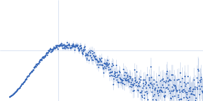
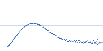
J-DNA binding domain monomer, 21 kDa Leishmania tarentolae protein
|
| Buffer: |
20 mM HEPES, 200 mM NaCl, 1 mM TCEP, pH: 7.5 |
| Experiment: |
SAXS
data collected at BM29, ESRF on 2017 Feb 4
|
The domain architecture of protozoan protein J-DNA-binding protein 1 suggests synergy between base J DNA binding and thymidine hydroxylase activity.
J Biol Chem (2019)
Adamopoulos A, Heidebrecht T, Roosendaal J, Touw WG, Phan IQ, Beijnen J, Perrakis A
|
| RgGuinier |
2.2 |
nm |
| Dmax |
7.1 |
nm |
| VolumePorod |
38 |
nm3 |
|
|
|
|
|
|
|
| Sample: |
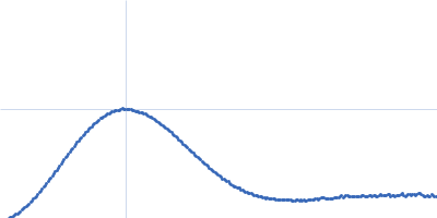
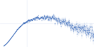
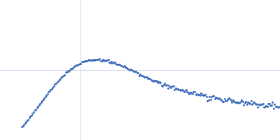
J-DNA (23mer) monomer, 14 kDa DNA
|
| Buffer: |
20 mM HEPES, 200 mM NaCl, 1 mM TCEP, pH: 7.5 |
| Experiment: |
SAXS
data collected at BM29, ESRF on 2016 Feb 21
|
The domain architecture of protozoan protein J-DNA-binding protein 1 suggests synergy between base J DNA binding and thymidine hydroxylase activity.
J Biol Chem (2019)
Adamopoulos A, Heidebrecht T, Roosendaal J, Touw WG, Phan IQ, Beijnen J, Perrakis A
|
| RgGuinier |
2.3 |
nm |
| Dmax |
7.3 |
nm |
| VolumePorod |
20 |
nm3 |
|
|
|
|
|
|
|
| Sample: |
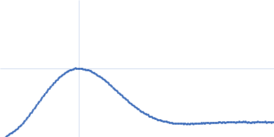
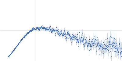
J-DNA binding domain monomer, 21 kDa Leishmania tarentolae protein
J-DNA (23mer) monomer, 14 kDa DNA
|
| Buffer: |
20 mM HEPES, 200 mM NaCl, 1 mM TCEP, pH: 7.5 |
| Experiment: |
SAXS
data collected at BM29, ESRF on 2016 Feb 21
|
The domain architecture of protozoan protein J-DNA-binding protein 1 suggests synergy between base J DNA binding and thymidine hydroxylase activity.
J Biol Chem (2019)
Adamopoulos A, Heidebrecht T, Roosendaal J, Touw WG, Phan IQ, Beijnen J, Perrakis A
|
| RgGuinier |
2.5 |
nm |
| Dmax |
8.6 |
nm |
| VolumePorod |
43 |
nm3 |
|
|
|
|
|
|
|
| Sample: |
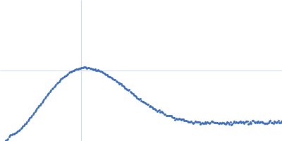
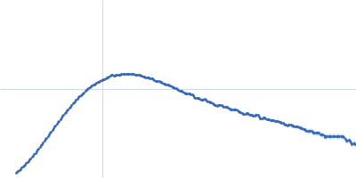
Thymine dioxygenase JBP1 monomer, 93 kDa Leishmania tarentolae protein
J-DNA (23mer) monomer, 14 kDa DNA
|
| Buffer: |
20 mM HEPES, 200 mM NaCl, 1 mM TCEP, pH: 7.5 |
| Experiment: |
SAXS
data collected at BM29, ESRF on 2016 Feb 21
|
The domain architecture of protozoan protein J-DNA-binding protein 1 suggests synergy between base J DNA binding and thymidine hydroxylase activity.
J Biol Chem (2019)
Adamopoulos A, Heidebrecht T, Roosendaal J, Touw WG, Phan IQ, Beijnen J, Perrakis A
|
| RgGuinier |
4.1 |
nm |
| Dmax |
14.1 |
nm |
| VolumePorod |
148 |
nm3 |
|
|
|
|
|
|
|
| Sample: |
Human Latent Transforming Growth Factor beta 1 dimer, 86 kDa Homo sapiens protein
|
| Buffer: |
phosphate buffered saline 2% glycerol, pH: 7.4 |
| Experiment: |
SAXS
data collected at 12.3.1 (SIBYLS), Advanced Light Source (ALS) on 2018 Oct 4
|
Structural consequences of transforming growth factor beta-1 activation from near-therapeutic X-ray doses.
J Synchrotron Radiat 26(Pt 4):967-979 (2019)
Stachowski T, Grant TD, Snell EH
|
| RgGuinier |
3.8 |
nm |
| Dmax |
17.5 |
nm |
| VolumePorod |
200 |
nm3 |
|
|
|
|
|
|
|
| Sample: |
Latency Associated Peptide dimer, 58 kDa Homo sapiens protein
|
| Buffer: |
phosphate buffered saline, pH: 7.4 |
| Experiment: |
SAXS
data collected at 12.3.1 (SIBYLS), Advanced Light Source (ALS) on 2018 Oct 4
|
Structural consequences of transforming growth factor beta-1 activation from near-therapeutic X-ray doses.
J Synchrotron Radiat 26(Pt 4):967-979 (2019)
Stachowski T, Grant TD, Snell EH
|
| RgGuinier |
4.1 |
nm |
| Dmax |
17.5 |
nm |
| VolumePorod |
179 |
nm3 |
|
|
|
|
|
|
|
| Sample: |
Insulin glargine (Lantus ®) hexamer, 36 kDa protein
|
| Buffer: |
Lantus Formulation (30 µg Zinc cloride, 2.7 mg m-Cresol, 20 mg glycerol 85%), pH: 4 |
| Experiment: |
SAXS
data collected at EMBL P12, PETRA III on 2017 May 23
|
The quaternary structure of insulin glargine and glulisine under formulation conditions.
Biophys Chem 253:106226 (2019)
Nagel N, Graewert MA, Gao M, Heyse W, Jeffries CM, Svergun D, Berchtold H
|
| RgGuinier |
1.8 |
nm |
| Dmax |
5.3 |
nm |
|
|
|
|
|
|
|
| Sample: |
Protein translocase subunit SecA dimer, 204 kDa Escherichia coli protein
|
| Buffer: |
20mM HEPES, 100mM NaCl, 1mM TCEP, pH: 8 |
| Experiment: |
SAXS
data collected at BM29, ESRF on 2016 Jul 18
|
The C-terminal tail of the bacterial translocation ATPase SecA modulates its activity.
Elife 8 (2019)
Jamshad M, Knowles TJ, White SA, Ward DG, Mohammed F, Rahman KF, Wynne M, Hughes GW, Kramer G, Bukau B, Huber D
|
| RgGuinier |
4.2 |
nm |
| Dmax |
14.9 |
nm |
| VolumePorod |
424 |
nm3 |
|
|
|
|
|
|
|
| Sample: |
Protein translocase subunit SecA dimer, 199 kDa Escherichia coli protein
|
| Buffer: |
20mM HEPES, 100mM NaCl, 1mM TCEP, pH: 8 |
| Experiment: |
SAXS
data collected at BM29, ESRF on 2016 Jul 18
|
The C-terminal tail of the bacterial translocation ATPase SecA modulates its activity.
Elife 8 (2019)
Jamshad M, Knowles TJ, White SA, Ward DG, Mohammed F, Rahman KF, Wynne M, Hughes GW, Kramer G, Bukau B, Huber D
|
| RgGuinier |
4.2 |
nm |
| Dmax |
14.8 |
nm |
| VolumePorod |
380 |
nm3 |
|
|
|
|
|
|
|
| Sample: |
Protein translocase subunit SecA dimer, 189 kDa Escherichia coli protein
|
| Buffer: |
20mM HEPES, 100mM NaCl, 1mM TCEP, pH: 8 |
| Experiment: |
SAXS
data collected at BM29, ESRF on 2016 Jul 18
|
The C-terminal tail of the bacterial translocation ATPase SecA modulates its activity.
Elife 8 (2019)
Jamshad M, Knowles TJ, White SA, Ward DG, Mohammed F, Rahman KF, Wynne M, Hughes GW, Kramer G, Bukau B, Huber D
|
| RgGuinier |
4.5 |
nm |
| Dmax |
15.7 |
nm |
| VolumePorod |
398 |
nm3 |
|
|

 experimental SAS data")
 experimental SAS data")
 experimental SAS data")


 experimental SAS data")