|
|
|
|
|
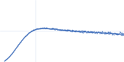
| Sample: |
DNA protection during starvation protein dodecamer, 224 kDa Escherichia coli (strain … protein
|
| Buffer: |
50 mM Tris-HCl, 50 mM NaCl, 0.5 mM EDTA, pH: 8 |
| Experiment: |
SAXS
data collected at EMBL P12, PETRA III on 2018 Nov 27
|
Protective Dps-DNA co-crystallization in stressed cells: an in vitro structural study by small-angle X-ray scattering and cryo-electron tomography.
FEBS Lett 593(12):1360-1371 (2019)
Dadinova LA, Chesnokov YM, Kamyshinsky RA, Orlov IA, Petoukhov MV, Mozhaev AA, Soshinskaya EY, Lazarev VN, Manuvera VA, Orekhov AS, Vasiliev AL, Shtykova EV
|
|
|
|
|
|
|
|
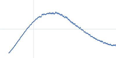
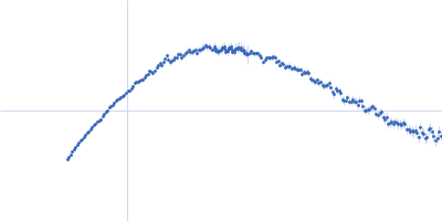
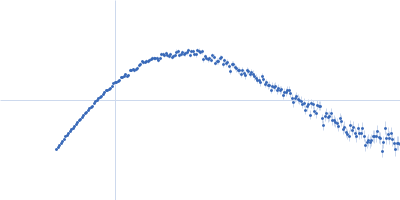
| Sample: |
Nuclear receptor CoRepressor 1; Nuclear Receptor Interaction Domain (NID) monomer, 29 kDa Mus musculus protein
Retinoid-X receptor alpha (RXR-alpha) Ligand Binding Domain (LBD) monomer, 26 kDa Mus musculus protein
Retinoic acid receptor alpha (RAR-alpha) Ligand binding domain (LDB) monomer, 28 kDa Homo sapiens protein
|
| Buffer: |
50 mM Tris-HCl, 150 mM NaCl, 2 mM TCEP, pH: 7.5 |
| Experiment: |
SAXS
data collected at BM29, ESRF on 2014 Jul 23
|
Interplay of Protein Disorder in Retinoic Acid Receptor Heterodimer and Its Corepressor Regulates Gene Expression.
Structure (2019)
Cordeiro TN, Sibille N, Germain P, Barthe P, Boulahtouf A, Allemand F, Bailly R, Vivat V, Ebel C, Barducci A, Bourguet W, le Maire A, Bernadó P
|
| RgGuinier |
4.8 |
nm |
| Dmax |
19.4 |
nm |
| VolumePorod |
167 |
nm3 |
|
|
|
|
|
|
|
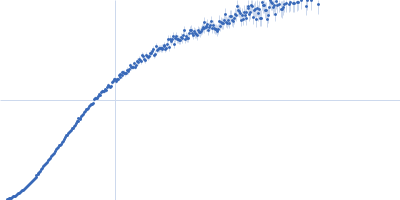
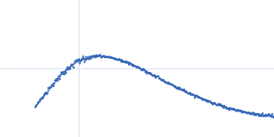
| Sample: |
Nuclear receptor CoRepressor 1; Nuclear Receptor Interaction Domain (NID) monomer, 29 kDa Mus musculus protein
Retinoid-X receptor alpha (RXR-alpha) Ligand Binding Domain (LBD) monomer, 26 kDa Mus musculus protein
Retinoid-X receptor alpha (RXR-alpha) Δ helix12 monomer, 24 kDa Mus musculus protein
|
| Buffer: |
50 mM Tris-HCl, 150 mM NaCl, 2 mM TCEP, pH: 7.5 |
| Experiment: |
SAXS
data collected at BM29, ESRF on 2014 Jul 23
|
Interplay of Protein Disorder in Retinoic Acid Receptor Heterodimer and Its Corepressor Regulates Gene Expression.
Structure (2019)
Cordeiro TN, Sibille N, Germain P, Barthe P, Boulahtouf A, Allemand F, Bailly R, Vivat V, Ebel C, Barducci A, Bourguet W, le Maire A, Bernadó P
|
| RgGuinier |
4.2 |
nm |
| Dmax |
15.7 |
nm |
| VolumePorod |
183 |
nm3 |
|
|
|
|
|
|
|
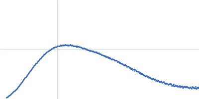
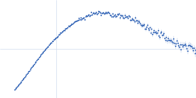
| Sample: |
Nuclear receptor CoRepressor 1; Nuclear Receptor Interaction Domain (NID) monomer, 29 kDa Mus musculus protein
Retinoid-X receptor alpha (RXR-alpha) Ligand Binding Domain (LBD) monomer, 26 kDa Mus musculus protein
Retinoic acid receptor alpha (RAR-alpha) Ligand binding domain (LDB) monomer, 28 kDa Homo sapiens protein
|
| Buffer: |
50 mM Tris-HCl, 150 mM NaCl, 2 mM TCEP, pH: 7.5 |
| Experiment: |
SAXS
data collected at BM29, ESRF on 2014 Jul 23
|
Interplay of Protein Disorder in Retinoic Acid Receptor Heterodimer and Its Corepressor Regulates Gene Expression.
Structure (2019)
Cordeiro TN, Sibille N, Germain P, Barthe P, Boulahtouf A, Allemand F, Bailly R, Vivat V, Ebel C, Barducci A, Bourguet W, le Maire A, Bernadó P
|
| RgGuinier |
4.8 |
nm |
| Dmax |
19.5 |
nm |
| VolumePorod |
178 |
nm3 |
|
|
|
|
|
|
|
| Sample: |
Nuclear receptor CoRepressor 1; Nuclear Receptor Interaction Domain (NID) monomer, 29 kDa Mus musculus protein
Retinoid-X receptor alpha (RXR-alpha) Ligand Binding Domain (LBD) monomer, 26 kDa Mus musculus protein
Retinoic acid receptor alpha (RAR-alpha) Ligand binding domain (LDB) mutant I396E monomer, 28 kDa Homo sapiens protein
|
| Buffer: |
50 mM Tris-HCl, 150 mM NaCl, 2 mM TCEP, pH: 7.5 |
| Experiment: |
SAXS
data collected at BM29, ESRF on 2015 Mar 9
|
Interplay of Protein Disorder in Retinoic Acid Receptor Heterodimer and Its Corepressor Regulates Gene Expression.
Structure (2019)
Cordeiro TN, Sibille N, Germain P, Barthe P, Boulahtouf A, Allemand F, Bailly R, Vivat V, Ebel C, Barducci A, Bourguet W, le Maire A, Bernadó P
|
| RgGuinier |
5.3 |
nm |
| Dmax |
22.4 |
nm |
| VolumePorod |
171 |
nm3 |
|
|
|
|
|
|
|
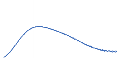
| Sample: |
Nuclear receptor CoRepressor 1; Nuclear Receptor Interaction Domain (NID) monomer, 29 kDa Mus musculus protein
Retinoid-X receptor alpha (RXR-alpha) Ligand Binding Domain (LBD) monomer, 26 kDa Mus musculus protein
Retinoic acid receptor alpha (RAR-alpha) Ligand binding domain (LDB) monomer, 28 kDa Homo sapiens protein
|
| Buffer: |
50 mM Tris-HCl, 150 mM NaCl, 2 mM TCEP, pH: 7.5 |
| Experiment: |
SAXS
data collected at BM29, ESRF on 2014 Jul 23
|
Interplay of Protein Disorder in Retinoic Acid Receptor Heterodimer and Its Corepressor Regulates Gene Expression.
Structure (2019)
Cordeiro TN, Sibille N, Germain P, Barthe P, Boulahtouf A, Allemand F, Bailly R, Vivat V, Ebel C, Barducci A, Bourguet W, le Maire A, Bernadó P
|
| RgGuinier |
4.2 |
nm |
| Dmax |
17.2 |
nm |
| VolumePorod |
131 |
nm3 |
|
|
|
|
|
|
|
| Sample: |
Resistance to inhibitors of cholinesterase 8 homolog A monomer, 51 kDa Rattus norvegicus protein
|
| Buffer: |
25 mM HEPES, 150 mM NaCl, pH: 8 |
| Experiment: |
SAXS
data collected at BL4-2, Stanford Synchrotron Radiation Lightsource (SSRL) on 2018 Apr 24
|
Structure, Function, and Dynamics of the Gα Binding Domain of Ric-8A.
Structure (2019)
Zeng B, Mou TC, Doukov TI, Steiner A, Yu W, Papasergi-Scott M, Tall GG, Hagn F, Sprang SR
|
| RgGuinier |
3.0 |
nm |
| Dmax |
10.6 |
nm |
| VolumePorod |
70 |
nm3 |
|
|
|
|
|
|
|
| Sample: |
Resistance to inhibitors of cholinesterase 8 homolog A monomer, 51 kDa Rattus norvegicus protein
|
| Buffer: |
25 mM HEPES, 150 mM NaCl, pH: 8 |
| Experiment: |
SAXS
data collected at BL4-2, Stanford Synchrotron Radiation Lightsource (SSRL) on 2018 Apr 24
|
Structure, Function, and Dynamics of the Gα Binding Domain of Ric-8A.
Structure (2019)
Zeng B, Mou TC, Doukov TI, Steiner A, Yu W, Papasergi-Scott M, Tall GG, Hagn F, Sprang SR
|
| RgGuinier |
3.0 |
nm |
| Dmax |
10.1 |
nm |
| VolumePorod |
70 |
nm3 |
|
|
|
|
|
|
|
| Sample: |
Dosage compensation regulator monomer, 29 kDa Drosophila melanogaster protein
RoX2 stem-loop 7, 18-mer fragment monomer, 12 kDa synthetic construct RNA
|
| Buffer: |
20 mM NaPO4, 200 mM NaCl, 1 mM DTT, pH: 6.5 |
| Experiment: |
SAXS
data collected at BM29, ESRF on 2016 Nov 29
|
Structure, dynamics and roX2-lncRNA binding of tandem double-stranded RNA binding domains dsRBD1,2 of Drosophila helicase Maleless.
Nucleic Acids Res 47(8):4319-4333 (2019)
Ankush Jagtap PK, Müller M, Masiewicz P, von Bülow S, Hollmann NM, Chen PC, Simon B, Thomae AW, Becker PB, Hennig J
|
| RgGuinier |
3.1 |
nm |
| Dmax |
13.3 |
nm |
| VolumePorod |
25 |
nm3 |
|
|
|
|
|
|
|
| Sample: |
Apolipoprotein E2 tetramer, 139 kDa Homo sapiens protein
|
| Buffer: |
20 mM HEPES, 300 mM NaCl, pH: 8 |
| Experiment: |
SAXS
data collected at B21, Diamond Light Source on 2017 Nov 29
|
The molecular basis for Apolipoprotein E4 as the major risk factor for late onset Alzheimer's disease.
J Mol Biol (2019)
Raulin AC, Kraft L, Al-Hilaly YK, Xue WF, McGeehan JE, Atack JR, Serpell L
|
| RgGuinier |
5.6 |
nm |
| Dmax |
19.5 |
nm |
| VolumePorod |
400 |
nm3 |
|
|

Retinoid-X receptor alpha (RXR-alpha) Ligand Binding Domain (LBD)Retinoic acid receptor alpha (RAR-alpha) Ligand binding domain (LDB) experimental SAS data")
Retinoid-X receptor alpha (RXR-alpha) Ligand Binding Domain (LBD)Retinoid-X receptor alpha (RXR-alpha) Δ helix12 experimental SAS data")
Retinoid-X receptor alpha (RXR-alpha) Ligand Binding Domain (LBD)Retinoic acid receptor alpha (RAR-alpha) Ligand binding domain (LDB) experimental SAS data")
Retinoid-X receptor alpha (RXR-alpha) Ligand Binding Domain (LBD)Retinoic acid receptor alpha (RAR-alpha) Ligand binding domain (LDB) mutant I396E experimental SAS data")
Retinoid-X receptor alpha (RXR-alpha) Ligand Binding Domain (LBD)Retinoic acid receptor alpha (RAR-alpha) Ligand binding domain (LDB) experimental SAS data")