|
|
|
|
|
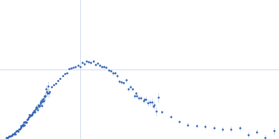
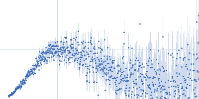
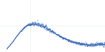
| Sample: |
1,2-dimyristoyl-sn-glycero-3-phosphocholine, 92 kDa
Apolipoprotein A-I dimer, 52 kDa Mus musculus protein
|
| Buffer: |
PBS in D2O, pH: 7.4 |
| Experiment: |
SANS
data collected at NG7, NIST Center for High Resolution Neutron Scattering (CHRNS) on 2015 Nov 25
|
Small-angle X-ray and neutron scattering demonstrates that cell-free expression produces properly formed disc-shaped nanolipoprotein particles.
Protein Sci 27(3):780-789 (2018)
Cleveland TE 4th, He W, Evans AC, Fischer NO, Lau EY, Coleman MA, Butler P
|
| RgGuinier |
3.5 |
nm |
| Dmax |
12.5 |
nm |
|
|
|
|
|
|
|
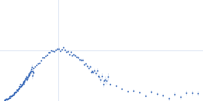
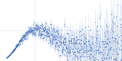
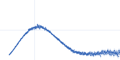
| Sample: |
1,2-dimyristoyl-sn-glycero-3-phosphocholine, 92 kDa
Apolipoprotein A-I dimer, 52 kDa Mus musculus protein
|
| Buffer: |
PBS in D2O, pH: 7.4 |
| Experiment: |
SANS
data collected at NG7, NIST Center for High Resolution Neutron Scattering (CHRNS) on 2015 Nov 25
|
Small-angle X-ray and neutron scattering demonstrates that cell-free expression produces properly formed disc-shaped nanolipoprotein particles.
Protein Sci 27(3):780-789 (2018)
Cleveland TE 4th, He W, Evans AC, Fischer NO, Lau EY, Coleman MA, Butler P
|
| RgGuinier |
3.3 |
nm |
| Dmax |
10.0 |
nm |
|
|
|
|
|
|
|
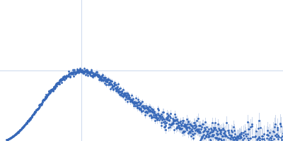
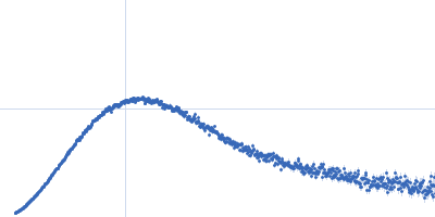
| Sample: |
EspG3 chaperone from Mycobacterium marinum M monomer, 32 kDa Mycobacterium marinum M protein
|
| Buffer: |
20 mM HEPES pH 7.5, 150 mM NaCl, pH: 7.5 |
| Experiment: |
SAXS
data collected at EMBL P12, PETRA III on 2014 Mar 17
|
Structural variability of EspG chaperones from mycobacterial ESX-1, ESX-3 and ESX-5 type VII secretion systems
(2018)
Tuukkanen A, Freire D, Chan S, Arbing M, Reed R, Evans T, Zenkeviciutė G, Kim J, Kahng S, Sawaya M, Chaton C, Wilmanns M, Eisenberg D, Parret A, Korotkov K
|
| RgGuinier |
2.3 |
nm |
| Dmax |
8.0 |
nm |
|
|
|
|
|
|
|
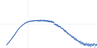
| Sample: |
EspG1 from Mycobacterium marinum monomer, 30 kDa Mycobacterium marinum protein
|
| Buffer: |
20 mM HEPES pH 7.5, 150 mM NaCl, pH: 7.5 |
| Experiment: |
SAXS
data collected at EMBL P12, PETRA III on 2014 Mar 17
|
Structural variability of EspG chaperones from mycobacterial ESX-1, ESX-3 and ESX-5 type VII secretion systems
(2018)
Tuukkanen A, Freire D, Chan S, Arbing M, Reed R, Evans T, Zenkeviciutė G, Kim J, Kahng S, Sawaya M, Chaton C, Wilmanns M, Eisenberg D, Parret A, Korotkov K
|
| RgGuinier |
2.7 |
nm |
| Dmax |
9.7 |
nm |
|
|
|
|
|
|
|
| Sample: |
EspG3 chaperone from Mycobacterium smegmatis monomer, 32 kDa Mycobacterium smegmatis protein
|
| Buffer: |
20 mM HEPES pH 7.5, 150 mM NaCl, pH: 7.5 |
| Experiment: |
SAXS
data collected at EMBL P12, PETRA III on 2014 Mar 17
|
Structural variability of EspG chaperones from mycobacterial ESX-1, ESX-3 and ESX-5 type VII secretion systems
(2018)
Tuukkanen A, Freire D, Chan S, Arbing M, Reed R, Evans T, Zenkeviciutė G, Kim J, Kahng S, Sawaya M, Chaton C, Wilmanns M, Eisenberg D, Parret A, Korotkov K
|
| RgGuinier |
2.5 |
nm |
| Dmax |
8.6 |
nm |
|
|
|
|
|
|
|
| Sample: |
EspG3 chaperone from Mycobacterium tuberculosis, 34 kDa Mycobacterium tuberculosis protein
|
| Buffer: |
20 mM HEPES pH 7.5, 150 mM NaCl, pH: 7.5 |
| Experiment: |
SAXS
data collected at EMBL P12, PETRA III on 2014 Mar 17
|
Structural variability of EspG chaperones from mycobacterial ESX-1, ESX-3 and ESX-5 type VII secretion systems
(2018)
Tuukkanen A, Freire D, Chan S, Arbing M, Reed R, Evans T, Zenkeviciutė G, Kim J, Kahng S, Sawaya M, Chaton C, Wilmanns M, Eisenberg D, Parret A, Korotkov K
|
| RgGuinier |
2.5 |
nm |
| Dmax |
9.0 |
nm |
|
|
|
|
|
|
|
| Sample: |
EspG3 chaperone from Mycobacterium smegmatis monomer, 32 kDa Mycobacterium smegmatis protein
|
| Buffer: |
20 mM HEPES pH 7.5, 150 mM NaCl, pH: 7.5 |
| Experiment: |
SAXS
data collected at EMBL P12, PETRA III on 2014 Mar 17
|
Structural variability of EspG chaperones from mycobacterial ESX-1, ESX-3 and ESX-5 type VII secretion systems
(2018)
Tuukkanen A, Freire D, Chan S, Arbing M, Reed R, Evans T, Zenkeviciutė G, Kim J, Kahng S, Sawaya M, Chaton C, Wilmanns M, Eisenberg D, Parret A, Korotkov K
|
| RgGuinier |
2.6 |
nm |
| Dmax |
9.2 |
nm |
|
|
|
|
|
|
|
| Sample: |
Polyubiquitin-C dimer, 17 kDa Homo sapiens protein
|
| Buffer: |
20mM HEPES, 150mM NaCl, pH: 7.4 |
| Experiment: |
SAXS
data collected at BL19U2, Shanghai Synchrotron Radiation Facility (SSRF) on 2016 Mar 24
|
Characterizing Protein Dynamics with Integrative Use of Bulk and Single-Molecule Techniques.
Biochemistry 57(3):305-313 (2018)
Liu Z, Gong Z, Cao Y, Ding YH, Dong MQ, Lu YB, Zhang WP, Tang C
|
| RgGuinier |
2.0 |
nm |
| Dmax |
7.0 |
nm |
| VolumePorod |
22 |
nm3 |
|
|
|
|
|
|
|
| Sample: |
Ammonium transporter histidine kinase domain monomer, 30 kDa Candidatus Kuenenia stuttgartiensis protein
|
| Buffer: |
20 mM Tris/HCl,100 mM NaCl, 5%(w/v) glycerol, pH: 8 |
| Experiment: |
SAXS
data collected at EMBL P12, PETRA III on 2013 Mar 2
|
Signaling ammonium across membranes through an ammonium sensor histidine kinase.
Nat Commun 9(1):164 (2018)
Pflüger T, Hernández CF, Lewe P, Frank F, Mertens H, Svergun D, Baumstark MW, Lunin VY, Jetten MSM, Andrade SLA
|
| RgGuinier |
2.7 |
nm |
| Dmax |
10.0 |
nm |
| VolumePorod |
71 |
nm3 |
|
|
|
|
|
|
|
| Sample: |
Ammonium transporter histidine kinase domain monomer, 30 kDa Candidatus Kuenenia stuttgartiensis protein
|
| Buffer: |
20 mM Tris/HCl,100 mM NaCl, 5%(w/v) glycerol, pH: 8 |
| Experiment: |
SAXS
data collected at EMBL P12, PETRA III on 2013 Mar 2
|
Signaling ammonium across membranes through an ammonium sensor histidine kinase.
Nat Commun 9(1):164 (2018)
Pflüger T, Hernández CF, Lewe P, Frank F, Mertens H, Svergun D, Baumstark MW, Lunin VY, Jetten MSM, Andrade SLA
|
| RgGuinier |
2.3 |
nm |
| Dmax |
8.0 |
nm |
| VolumePorod |
53 |
nm3 |
|
|