|
|
|
|
|
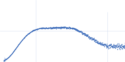
| Sample: |
TnpA transposase dimer, 234 kDa Bacillus thuringiensis serovar … protein
|
| Buffer: |
50 mM HEPES, 200 mM NaCl, 100 mM L-Arg HCL, pH: 7.9 |
| Experiment: |
SAXS
data collected at SWING, SOLEIL on 2017 Nov 2
|
AFM-based force spectroscopy unravels stepwise formation of the DNA transposition complex in the widespread Tn3 family mobile genetic elements.
Nucleic Acids Res (2023)
Fernandez M, Shkumatov AV, Liu Y, Stulemeijer C, Derclaye S, Efremov RG, Hallet B, Alsteens D
|
| RgGuinier |
4.6 |
nm |
| Dmax |
16.0 |
nm |
| VolumePorod |
480 |
nm3 |
|
|
|
|
|
|
|
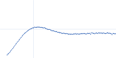
| Sample: |
Cell division protein FtsB hexamer, 70 kDa Escherichia coli (strain … protein
Cell division protein FtsL hexamer, 82 kDa Escherichia coli (strain … protein
Cell division protein FtsQ hexamer, 189 kDa Escherichia coli (strain … protein
|
| Buffer: |
20 mM HEPES pH 7.5, 200 mM NaCl, 0.2% of Cymal-5, pH: 7.5 |
| Experiment: |
SAXS
data collected at 13A, Taiwan Photon Source, NSRRC on 2022 Sep 23
|
Structure of the heterotrimeric membrane protein complex FtsB-FtsL-FtsQ of the bacterial divisome
Nature Communications 14(1) (2023)
Nguyen H, Chen X, Parada C, Luo A, Shih O, Jeng U, Huang C, Shih Y, Ma C
|
| RgGuinier |
5.3 |
nm |
| Dmax |
16.5 |
nm |
| VolumePorod |
421 |
nm3 |
|
|
|
|
|
|
|
| Sample: |
Isoform P3 of Phosphoprotein dimer, 55 kDa Rabies virus (strain … protein
|
| Buffer: |
25 mM HEPES, 150 mM NaCl, 1 mM TCEP, pH: 7.4 |
| Experiment: |
SAXS
data collected at SAXS/WAXS, Australian Synchrotron on 2019 Jun 26
|
Structural insights into the multifunctionality of rabies virus P3 protein.
Proc Natl Acad Sci U S A 120(14):e2217066120 (2023)
Sethi A, Rawlinson SM, Dubey A, Ang CS, Choi YH, Yan F, Okada K, Rozario AM, Brice AM, Ito N, Williamson NA, Hatters DM, Bell TDM, Arthanari H, Moseley GW, Gooley PR
|
| RgGuinier |
3.7 |
nm |
| Dmax |
16.5 |
nm |
| VolumePorod |
108 |
nm3 |
|
|
|
|
|
|
|
| Sample: |
Attenuated derivative P3 of Phosphoprotein dimer, 55 kDa protein
|
| Buffer: |
25 mM HEPES, 150 mM NaCl, 1 mM TCEP, pH: 7.4 |
| Experiment: |
SAXS
data collected at SAXS/WAXS, Australian Synchrotron on 2019 Jun 26
|
Structural insights into the multifunctionality of rabies virus P3 protein.
Proc Natl Acad Sci U S A 120(14):e2217066120 (2023)
Sethi A, Rawlinson SM, Dubey A, Ang CS, Choi YH, Yan F, Okada K, Rozario AM, Brice AM, Ito N, Williamson NA, Hatters DM, Bell TDM, Arthanari H, Moseley GW, Gooley PR
|
| RgGuinier |
4.0 |
nm |
| Dmax |
17.5 |
nm |
| VolumePorod |
127 |
nm3 |
|
|
|
|
|
|
|
| Sample: |
Isoform P3 of Phosphoprotein Nish P3 N226H dimer, 55 kDa protein
|
| Buffer: |
25 mM HEPES, 150 mM NaCl, 1 mM TCEP, pH: 7.4 |
| Experiment: |
SAXS
data collected at SAXS/WAXS, Australian Synchrotron on 2019 Jun 26
|
Structural insights into the multifunctionality of rabies virus P3 protein.
Proc Natl Acad Sci U S A 120(14):e2217066120 (2023)
Sethi A, Rawlinson SM, Dubey A, Ang CS, Choi YH, Yan F, Okada K, Rozario AM, Brice AM, Ito N, Williamson NA, Hatters DM, Bell TDM, Arthanari H, Moseley GW, Gooley PR
|
| RgGuinier |
4.3 |
nm |
| Dmax |
19.0 |
nm |
| VolumePorod |
150 |
nm3 |
|
|
|
|
|
|
|
| Sample: |
Calmodulin-1 monomer, 17 kDa Homo sapiens protein
|
| Buffer: |
50 mM HEPES, 100 mM NaCl, 2 mM CaCl2, 1 mM TCEP, pH: 7.4 |
| Experiment: |
SAXS
data collected at 12.3.1 (SIBYLS), Advanced Light Source (ALS) on 2021 Sep 23
|
Binding by calmodulin is coupled to transient unfolding of the third FF domain of Prp40A.
Protein Sci 32(4):e4606 (2023)
Díaz Casas A, Cordoba JJ, Ferrer BJ, Balakrishnan S, Wurm JE, Pastrana-Ríos B, Chazin WJ
|
| RgGuinier |
2.2 |
nm |
| Dmax |
7.0 |
nm |
| VolumePorod |
38 |
nm3 |
|
|
|
|
|
|
|
| Sample: |
Pre-mRNA-processing factor 40 homolog A monomer, 9 kDa Homo sapiens protein
|
| Buffer: |
50 mM HEPES, 100 mM NaCl, 2 mM CaCl2, 1 mM TCEP, pH: 7.4 |
| Experiment: |
SAXS
data collected at 12.3.1 (SIBYLS), Advanced Light Source (ALS) on 2021 Sep 23
|
Binding by calmodulin is coupled to transient unfolding of the third FF domain of Prp40A.
Protein Sci 32(4):e4606 (2023)
Díaz Casas A, Cordoba JJ, Ferrer BJ, Balakrishnan S, Wurm JE, Pastrana-Ríos B, Chazin WJ
|
| RgGuinier |
1.6 |
nm |
| Dmax |
7.2 |
nm |
| VolumePorod |
13 |
nm3 |
|
|
|
|
|
|
|
| Sample: |
Calmodulin-1 monomer, 17 kDa Homo sapiens protein
Pre-mRNA-processing factor 40 homolog A monomer, 9 kDa Homo sapiens protein
|
| Buffer: |
50 mM HEPES, 100 mM NaCl, 2 mM CaCl2, 1 mM TCEP, pH: 7.4 |
| Experiment: |
SAXS
data collected at 12.3.1 (SIBYLS), Advanced Light Source (ALS) on 2021 Sep 23
|
Binding by calmodulin is coupled to transient unfolding of the third FF domain of Prp40A.
Protein Sci 32(4):e4606 (2023)
Díaz Casas A, Cordoba JJ, Ferrer BJ, Balakrishnan S, Wurm JE, Pastrana-Ríos B, Chazin WJ
|
| RgGuinier |
2.4 |
nm |
| Dmax |
8.7 |
nm |
| VolumePorod |
29 |
nm3 |
|
|
|
|
|
|
|
| Sample: |
Colibactin hybrid non-ribosomal peptide synthetase/type I polyketide synthase ClbK dimer, 476 kDa Escherichia coli protein
|
| Buffer: |
50 mM Hepes pH 7.5, 200 mM NaCl, 5 mM DTT, 5 mM MgCl2, pH: 7.5 |
| Experiment: |
SAXS
data collected at BM29, ESRF on 2020 Sep 14
|
Architecture of a PKS-NRPS hybrid megaenzyme involved in the biosynthesis of the genotoxin colibactin
Structure (2023)
Bonhomme S, Contreras-Martel C, Dessen A, Macheboeuf P
|
| RgGuinier |
8.0 |
nm |
| Dmax |
30.0 |
nm |
| VolumePorod |
1340 |
nm3 |
|
|
|
|
|
|
|
| Sample: |
Human dystrophin central domain R4-15 fragment monomer, 150 kDa protein
|
| Buffer: |
NaP 20 mM, NaCl 300 mM, EDTA 1 mM, Glycérol 2%, pH: 7.5 |
| Experiment: |
SAXS
data collected at SWING, SOLEIL on 2016 May 26
|
Dystrophin SAXS data
Raphael Dos Santos Morais
|
| RgGuinier |
9.7 |
nm |
| Dmax |
47.5 |
nm |
|
|


 Rg histogram")

 Rg histogram")

 Rg histogram")