|
|
|
|
|
| Sample: |
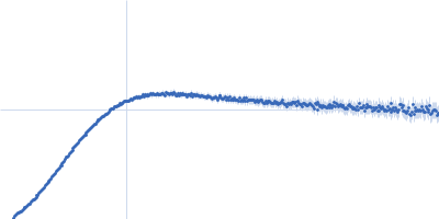
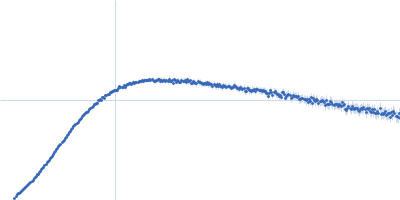
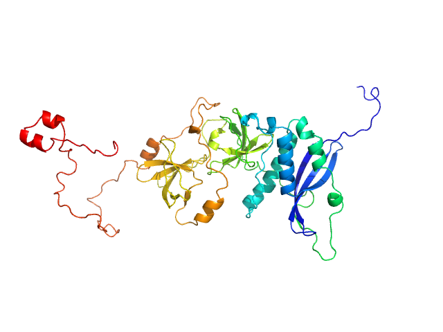
Neutophil cytosol factor 1 monomer, 40 kDa Homo sapiens protein
|
| Buffer: |
50 mM HEPES, 100 mM NaCl, 1 mM EDTA, 2 mM DTT, 5% glycerol, pH: 7.5 |
| Experiment: |
SAXS
data collected at Bruker Nanostar, IBBMC on 2009 Oct 16
|
Quantitative live-cell imaging and 3D modeling reveal critical functional features in the cytosolic complex of phagocyte NADPH oxidase.
J Biol Chem (2019)
Ziegler CS, Bouchab L, Tramier M, Durand D, Fieschi F, Dupré-Crochet S, Mérola F, Nüße O, Erard M
|
| RgGuinier |
2.6 |
nm |
| Dmax |
10.0 |
nm |
| VolumePorod |
58 |
nm3 |
|
|
|
|
|
|
|
| Sample: |
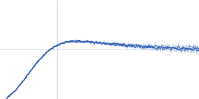
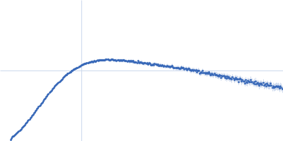
Neutrophil cytosol factor 1 monomer, 46 kDa Homo sapiens protein
|
| Buffer: |
50 mM HEPES, 100 mM NaCl, 1 mM EDTA, 2 mM DTT, 5% glycerol, pH: 7.5 |
| Experiment: |
SAXS
data collected at SWING, SOLEIL on 2008 Apr 23
|
Quantitative live-cell imaging and 3D modeling reveal critical functional features in the cytosolic complex of phagocyte NADPH oxidase.
J Biol Chem (2019)
Ziegler CS, Bouchab L, Tramier M, Durand D, Fieschi F, Dupré-Crochet S, Mérola F, Nüße O, Erard M
|
| RgGuinier |
3.2 |
nm |
| Dmax |
12.5 |
nm |
| VolumePorod |
77 |
nm3 |
|
|
|
|
|
|
|
| Sample: |
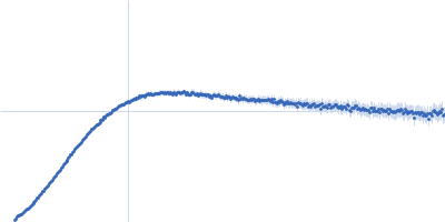
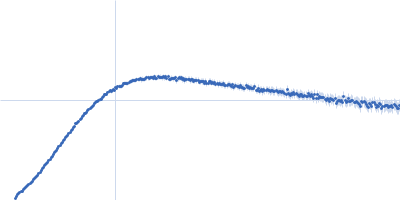
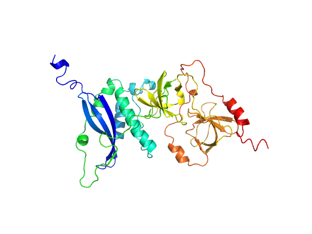
Neutrophil cytosol factor 2 monomer, 61 kDa Homo sapiens protein
|
| Buffer: |
20 mM HEPES, 50 mM NaCl, 1 mM EDTA, 2 mM DTT, 5% glycerol, pH: 8 |
| Experiment: |
SAXS
data collected at D24, LURE on 2003 Apr 9
|
Quantitative live-cell imaging and 3D modeling reveal critical functional features in the cytosolic complex of phagocyte NADPH oxidase.
J Biol Chem (2019)
Ziegler CS, Bouchab L, Tramier M, Durand D, Fieschi F, Dupré-Crochet S, Mérola F, Nüße O, Erard M
|
| RgGuinier |
4.3 |
nm |
| Dmax |
16.0 |
nm |
| VolumePorod |
113 |
nm3 |
|
|
|
|
|
|
|
| Sample: |
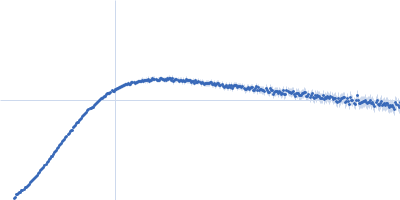
Apolipoprotein E2 tetramer, 139 kDa Homo sapiens protein
Heparin monomer, 15 kDa
|
| Buffer: |
20 mM HEPES, 300 mM NaCl, 1 mM TCEP, pH: 8 |
| Experiment: |
SAXS
data collected at B21, Diamond Light Source on 2018 May 3
|
Therapeutic approaches to ApoE
University of Sussex PhD thesis 2019 (2019)
Lucas Kraft
|
| RgGuinier |
6.2 |
nm |
| Dmax |
20.7 |
nm |
| VolumePorod |
504 |
nm3 |
|
|
|
|
|
|
|
| Sample: |
Apolipoprotein E2 tetramer, 139 kDa Homo sapiens protein
Heparin monomer, 15 kDa
|
| Buffer: |
20 mM HEPES, 300 mM NaCl, 1 mM TCEP, pH: 8 |
| Experiment: |
SAXS
data collected at B21, Diamond Light Source on 2018 May 3
|
Therapeutic approaches to ApoE
University of Sussex PhD thesis 2019 (2019)
Lucas Kraft
|
| RgGuinier |
6.4 |
nm |
| Dmax |
21.7 |
nm |
| VolumePorod |
531 |
nm3 |
|
|
|
|
|
|
|
| Sample: |
Apolipoprotein E2 tetramer, 139 kDa Homo sapiens protein
Heparin monomer, 15 kDa
|
| Buffer: |
20 mM HEPES, 300 mM NaCl, 1 mM TCEP, pH: 8 |
| Experiment: |
SAXS
data collected at B21, Diamond Light Source on 2018 May 3
|
Therapeutic approaches to ApoE
University of Sussex PhD thesis 2019 (2019)
Lucas Kraft
|
| RgGuinier |
6.5 |
nm |
| Dmax |
23.4 |
nm |
| VolumePorod |
584 |
nm3 |
|
|
|
|
|
|
|
| Sample: |
Apolipoprotein E2 tetramer, 139 kDa Homo sapiens protein
Heparin monomer, 15 kDa
|
| Buffer: |
20 mM HEPES, 300 mM NaCl, 1 mM TCEP, pH: 8 |
| Experiment: |
SAXS
data collected at B21, Diamond Light Source on 2018 May 3
|
Therapeutic approaches to ApoE
University of Sussex PhD thesis 2019 (2019)
Lucas Kraft
|
| RgGuinier |
7.1 |
nm |
| Dmax |
24.0 |
nm |
| VolumePorod |
666 |
nm3 |
|
|
|
|
|
|
|
| Sample: |
Apolipoprotein E2 tetramer, 139 kDa Homo sapiens protein
Heparin monomer, 15 kDa
|
| Buffer: |
20 mM HEPES, 300 mM NaCl, 1 mM TCEP, pH: 8 |
| Experiment: |
SAXS
data collected at B21, Diamond Light Source on 2018 May 3
|
Therapeutic approaches to ApoE
University of Sussex PhD thesis 2019 (2019)
Lucas Kraft
|
| RgGuinier |
7.2 |
nm |
| Dmax |
24.5 |
nm |
| VolumePorod |
696 |
nm3 |
|
|
|
|
|
|
|
| Sample: |
Apolipoprotein E3 tetramer, 139 kDa Homo sapiens protein
Heparin monomer, 15 kDa
|
| Buffer: |
20 mM HEPES, 300 mM NaCl, 1 mM TCEP, pH: 8 |
| Experiment: |
SAXS
data collected at B21, Diamond Light Source on 2018 May 3
|
Therapeutic approaches to ApoE
University of Sussex PhD thesis 2019 (2019)
Lucas Kraft
|
| RgGuinier |
7.3 |
nm |
| Dmax |
25.9 |
nm |
|
|
|
|
|
|
|
| Sample: |
Apolipoprotein E3 tetramer, 139 kDa Homo sapiens protein
Heparin monomer, 15 kDa
|
| Buffer: |
20 mM HEPES, 300 mM NaCl, 1 mM TCEP, pH: 8 |
| Experiment: |
SAXS
data collected at B21, Diamond Light Source on 2018 May 3
|
Therapeutic approaches to ApoE
University of Sussex PhD thesis 2019 (2019)
Lucas Kraft
|
| RgGuinier |
7.5 |
nm |
| Dmax |
25.8 |
nm |
|
|


 subunit of phagocyte NADPH oxidase Rg histogram")