|
|
|
|
|
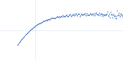
| Sample: |
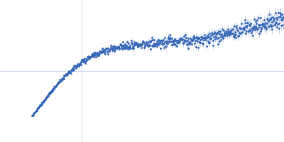
Heparan sulphate oligomer monomer, 20 kDa Sus scrofa domesticus
|
| Buffer: |
50 mM Tris-HCl, 120 mM CaCl2, pH: 7.4 |
| Experiment: |
SAXS
data collected at SWING, SOLEIL on 2020 Sep 13
|
Comparative analysis of heparine oligosaccharides, heparin and heparan sulphate
Adriana Erica Miele
|
| RgGuinier |
3.8 |
nm |
| Dmax |
20.6 |
nm |
|
|
|
|
|
|
|
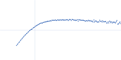
| Sample: |
Protein W monomer, 53 kDa Hendra virus (isolate … protein
|
| Buffer: |
20 mM HEPES, 150 mM NaCl, pH: 7.2 |
| Experiment: |
SAXS
data collected at SWING, SOLEIL on 2022 Jul 14
|
Henipavirus W proteins
Frank Gondelaud
|
| RgGuinier |
6.8 |
nm |
| Dmax |
26.0 |
nm |
| VolumePorod |
250 |
nm3 |
|
|
|
|
|
|
|
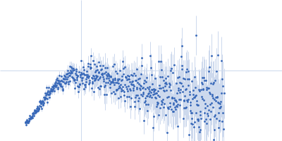
| Sample: |
Protein W monomer, 47 kDa Hendra virus (isolate … protein
|
| Buffer: |
20 mM HEPES, 150 mM NaCl, pH: 7.2 |
| Experiment: |
SAXS
data collected at SWING, SOLEIL on 2022 Jul 14
|
Henipavirus W proteins
Frank Gondelaud
|
| RgGuinier |
6.4 |
nm |
| Dmax |
32.0 |
nm |
|
|
|
|
|
|
|
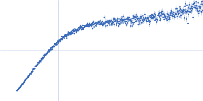
| Sample: |
Protein W mutant (C316S, C334S, C419S) dimer, 105 kDa Hendra virus (isolate … protein
|
| Buffer: |
20 mM HEPES, 150 mM NaCl, pH: 7.2 |
| Experiment: |
SAXS
data collected at SWING, SOLEIL on 2022 Jul 14
|
Henipavirus W proteins
Frank Gondelaud
|
| RgGuinier |
8.1 |
nm |
| Dmax |
37.0 |
nm |
|
|
|
|
|
|
|
| Sample: |
Genome polyprotein (Protein 2A H-NC; Δ130-150), 15 kDa Human parechovirus 1 … protein
|
| Buffer: |
20 mM HEPES, 150 mM NaCl, 5 mM DDT, pH: 7.4 |
| Experiment: |
SAXS
data collected at EMBL P12, PETRA III on 2018 May 28
|
Structural plasticity of 2A proteins in the Parechovirus family
Zuzanna Pietras
|
| RgGuinier |
2.0 |
nm |
| Dmax |
9.5 |
nm |
| VolumePorod |
43 |
nm3 |
|
|
|
|
|
|
|
| Sample: |
Pigeon iron-sulfur cluster assembly 1 homolog, mitochondrial, 15 kDa Columba livia protein
|
| Buffer: |
20 mM Tris-HCl, 0.15 M NaCl, 10 mM 3-mercapto-1,2-propanediol, pH: 8 |
| Experiment: |
SAXS
data collected at BL-10C, Photon Factory (PF), High Energy Accelerator Research Organization (KEK) on 2021 Jun 8
|
A hidden property of the iron-sulfur protein in the mononuclear iron-bound state: species-dependent structural ordering induced by magnetic fields.
FEBS J (2025)
Arai S, Soga S, Hirai M, Kobayashi R, Masai H, Kimura K, Maeda K, Nagashima H
|
| RgGuinier |
2.8 |
nm |
| Dmax |
10.5 |
nm |
| VolumePorod |
85 |
nm3 |
|
|
|
|
|
|
|
| Sample: |
Pigeon iron-sulfur cluster assembly 1 homolog, mitochondrial, 15 kDa Columba livia protein
|
| Buffer: |
20 mM Tris-HCl, 0.15 M NaCl, 10 mM 3-mercapto-1,2-propanediol, pH: 8 |
| Experiment: |
SAXS
data collected at BL-10C, Photon Factory (PF), High Energy Accelerator Research Organization (KEK) on 2021 Nov 1
|
A hidden property of the iron-sulfur protein in the mononuclear iron-bound state: species-dependent structural ordering induced by magnetic fields.
FEBS J (2025)
Arai S, Soga S, Hirai M, Kobayashi R, Masai H, Kimura K, Maeda K, Nagashima H
|
|
|
|
|
|
|
|
| Sample: |
Pigeon iron-sulfur cluster assembly 1 homolog, mitochondrial, 15 kDa Columba livia protein
|
| Buffer: |
20 mM Tris-HCl, 0.15 M NaCl, 10 mM 3-mercapto-1,2-propanediol, pH: 8 |
| Experiment: |
SAXS
data collected at BL-10C, Photon Factory (PF), High Energy Accelerator Research Organization (KEK) on 2021 Nov 1
|
A hidden property of the iron-sulfur protein in the mononuclear iron-bound state: species-dependent structural ordering induced by magnetic fields.
FEBS J (2025)
Arai S, Soga S, Hirai M, Kobayashi R, Masai H, Kimura K, Maeda K, Nagashima H
|
|
|
|
|
|
|

|
| Sample: |
Pigeon iron-sulfur cluster assembly 1 homolog, mitochondrial, 15 kDa Columba livia protein
|
| Buffer: |
20 mM Tris-HCl, 0.15 M NaCl, 10 mM 3-mercapto-1,2-propanediol, pH: 8 |
| Experiment: |
SAXS
data collected at BL-10C, Photon Factory (PF), High Energy Accelerator Research Organization (KEK) on 2021 Jun 8
|
A hidden property of the iron-sulfur protein in the mononuclear iron-bound state: species-dependent structural ordering induced by magnetic fields.
FEBS J (2025)
Arai S, Soga S, Hirai M, Kobayashi R, Masai H, Kimura K, Maeda K, Nagashima H
|
| RgGuinier |
2.7 |
nm |
| Dmax |
7.2 |
nm |
| VolumePorod |
39 |
nm3 |
|
|
|
|
|
|
|
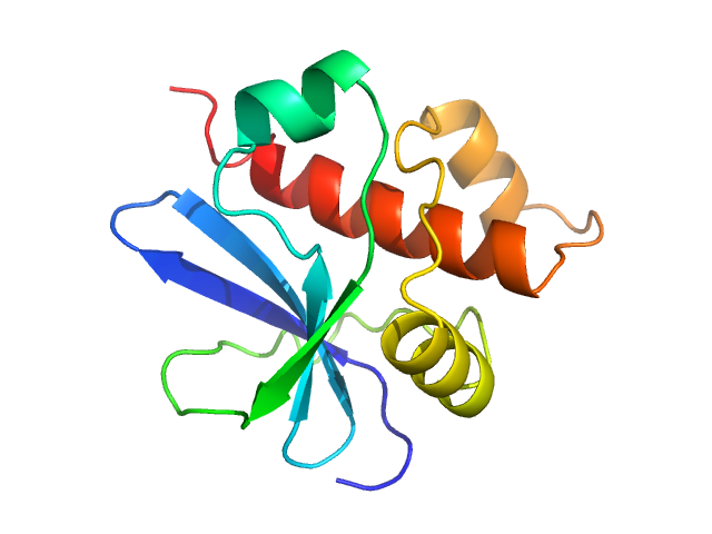
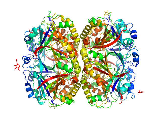
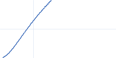
| Sample: |
Perivitellin ovorubin-1 dimer, 41 kDa Pomacea canaliculata protein
Perivitellin ovorubin-2 dimer, 42 kDa Pomacea canaliculata protein
Uncharacterized protein (ovorubin-3, short) dimer, 40 kDa Pomacea canaliculata protein
Perivitellin protein (ovorubin-4, form 1) dimer, 40 kDa Pomacea canaliculata protein
Uncharacterized protein (ovorubin-5) dimer, 41 kDa Pomacea canaliculata protein
|
| Buffer: |
20 mM Tris, 150 mM NaCl, pH: 7 |
| Experiment: |
SAXS
data collected at TPS13A, NSRRC on 2025 Mar 26
|
Structure of the chromoprotein ovorubin from the golden apple snail ( Pomacea canaliculata
)
Protein Science 35(1) (2025)
Wilasluck P, Saw W, Tran B, Sabat G, Shih O, Hengphasatporn K, Shigeta Y, Vinayavekhin N, Wangkanont K
|
| RgGuinier |
4.1 |
nm |
| Dmax |
12.4 |
nm |
| VolumePorod |
400 |
nm3 |
|
|


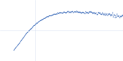

 experimental SAS data")
 experimental SAS data")


Perivitellin protein (ovorubin-4, form 1)Uncharacterized protein (ovorubin-5) experimental SAS data")