|
|
|
|
|
| Sample: |
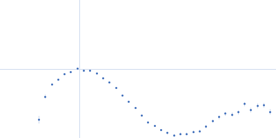
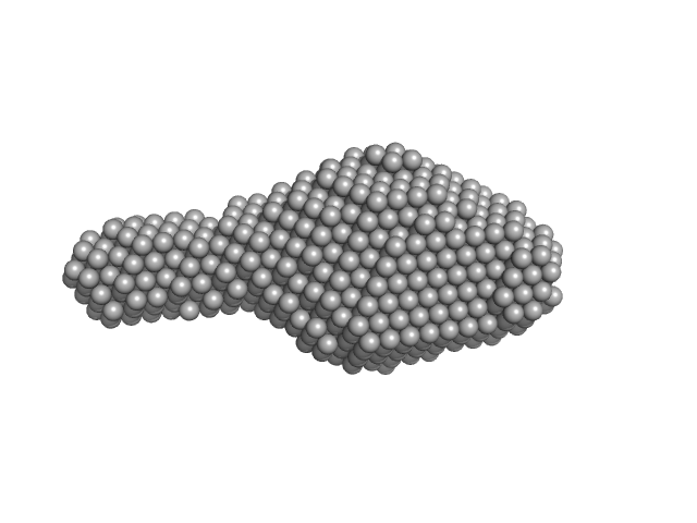
Collagenase ColG segement s2s3as3b monomer, 37 kDa Hathewaya histolytica protein
|
| Buffer: |
10mM HEPES 100mM NaCl 0.2mM EGTA, pH: 7.5 |
| Experiment: |
SAXS
data collected at 12.3.1 (SIBYLS), Advanced Light Source (ALS) on 2016 Oct 11
|
Ca2+ - Induced Structural Change of Multi-Domain Collagen Binding Segments of Collagenases ColG and ColH from Hathewaya histolytica
University of Arkansas Dissertation - (2018)
Christopher E Ruth
|
| RgGuinier |
3.0 |
nm |
| Dmax |
14.4 |
nm |
| VolumePorod |
61 |
nm3 |
|
|
|
|
|
|
|
| Sample: |
Human respiratory syncytial virus M2-1 monomer, 14 kDa Human orthopneumovirus protein
|
| Buffer: |
20 mM Tris–HCl, 300 mM NaCl,, pH: 7 |
| Experiment: |
SAXS
data collected at EMBL P12, PETRA III on 2016 Dec 13
|
Structure and stability of the Human respiratory syncytial virus M2-1 RNA-binding core domain reveals a compact and cooperative folding unit.
Acta Crystallogr F Struct Biol Commun 74(Pt 1):23-30 (2018)
Molina IG, Josts I, Almeida Hernandez Y, Esperante S, Salgueiro M, Garcia Alai MM, de Prat-Gay G, Tidow H
|
| RgGuinier |
2.0 |
nm |
| Dmax |
7.9 |
nm |
| VolumePorod |
3 |
nm3 |
|
|
|
|
|
|
|
| Sample: |
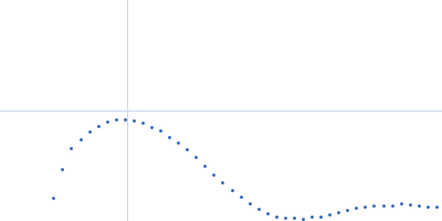
Sensory box protein light-state (R66I) dimer, 37 kDa Pseudomonas putida protein
|
| Buffer: |
10mM Tris, 10 mM NaCl, pH: 7 |
| Experiment: |
SAXS
data collected at BM29, ESRF on 2014 Dec 2
|
Small-angle X-ray scattering study of the kinetics of light-dark transition in a LOV protein.
PLoS One 13(7):e0200746 (2018)
Röllen K, Granzin J, Batra-Safferling R, Stadler AM
|
| RgGuinier |
2.6 |
nm |
| Dmax |
9.2 |
nm |
| VolumePorod |
58 |
nm3 |
|
|
|
|
|
|
|
| Sample: |
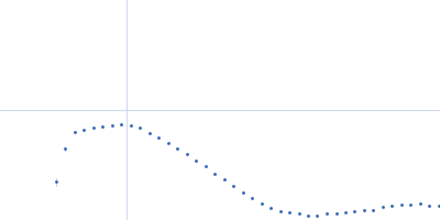
Sensory box protein dark-state dimer, 37 kDa Pseudomonas putida protein
|
| Buffer: |
10mM Tris, 10 mM NaCl, pH: 7 |
| Experiment: |
SAXS
data collected at BM29, ESRF on 2014 Dec 2
|
Small-angle X-ray scattering study of the kinetics of light-dark transition in a LOV protein.
PLoS One 13(7):e0200746 (2018)
Röllen K, Granzin J, Batra-Safferling R, Stadler AM
|
| RgGuinier |
2.6 |
nm |
| Dmax |
8.0 |
nm |
| VolumePorod |
57 |
nm3 |
|
|
|
|
|
|
|
| Sample: |
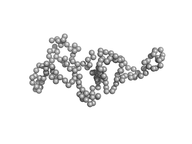
Disrupted- in-schizophrenia 1 (DISC1 12D2) 691-836 monomer, 19 kDa protein
|
| Buffer: |
25 mM Tris-HCl, 150 mM NaCl, 1mM DTT, pH: 7.4 |
| Experiment: |
SAXS
data collected at EMBL P12, PETRA III on 2015 Oct 1
|
Biophysical insights from a single chain camelid antibody directed against the Disrupted-in-Schizophrenia 1 protein.
PLoS One 13(1):e0191162 (2018)
Yerabham ASK, Müller-Schiffmann A, Ziehm T, Stadler A, Köber S, Indurkhya X, Marreiros R, Trossbach SV, Bradshaw NJ, Prikulis I, Willbold D, Weiergräber OH, Korth C
|
| RgGuinier |
2.6 |
nm |
| Dmax |
7.3 |
nm |
| VolumePorod |
41 |
nm3 |
|
|
|
|
|
|
|
| Sample: |
Anti-DISC1 single-domain camelid antibody VHH B5 monomer, 14 kDa protein
|
| Buffer: |
25 mM Tris, 150 mM NaCl, pH: 7.4 |
| Experiment: |
SAXS
data collected at BM29, ESRF on 2016 Feb 19
|
Biophysical insights from a single chain camelid antibody directed against the Disrupted-in-Schizophrenia 1 protein.
PLoS One 13(1):e0191162 (2018)
Yerabham ASK, Müller-Schiffmann A, Ziehm T, Stadler A, Köber S, Indurkhya X, Marreiros R, Trossbach SV, Bradshaw NJ, Prikulis I, Willbold D, Weiergräber OH, Korth C
|
| RgGuinier |
1.8 |
nm |
| Dmax |
8.4 |
nm |
| VolumePorod |
21 |
nm3 |
|
|
|
|
|
|
|
| Sample: |
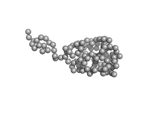
Anti-DISC1 single-domain camelid antibody VHH B5 monomer, 14 kDa protein
Disrupted- in-schizophrenia 1 (DISC1 12D2) 691-836 monomer, 19 kDa protein
|
| Buffer: |
25 mM Tris-HCl, 150 mM NaCl, 1mM DTT, pH: 7.4 |
| Experiment: |
SAXS
data collected at BM29, ESRF on 2016 Feb 19
|
Biophysical insights from a single chain camelid antibody directed against the Disrupted-in-Schizophrenia 1 protein.
PLoS One 13(1):e0191162 (2018)
Yerabham ASK, Müller-Schiffmann A, Ziehm T, Stadler A, Köber S, Indurkhya X, Marreiros R, Trossbach SV, Bradshaw NJ, Prikulis I, Willbold D, Weiergräber OH, Korth C
|
| RgGuinier |
3.1 |
nm |
| Dmax |
10.4 |
nm |
| VolumePorod |
77 |
nm3 |
|
|
|
|
|
|
|
| Sample: |
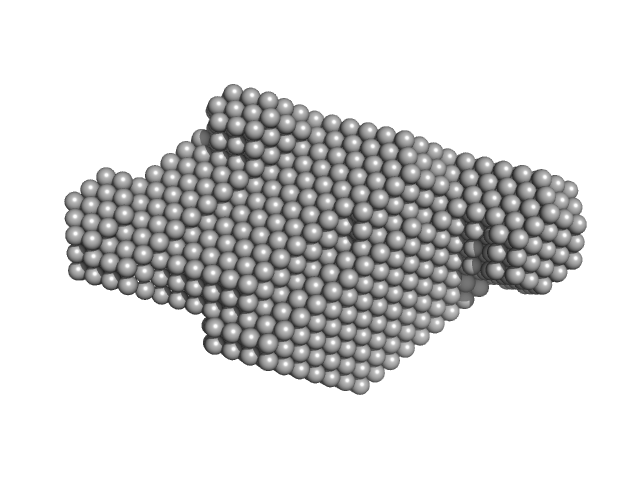
Bacteriophage phi-X174 monomer, 0 kDa protein
|
| Buffer: |
0.06 M NH4Cl2, 0.09 M NaCl, 0.1 M KCl, 1 mM MgS04, 1 mM CaCl2, 0.1 M Tris-HCl, pH: 7.4 |
| Experiment: |
SAXS
data collected at G1, Cornell High Energy Synchrotron Source (CHESS) on 2015 Oct 25
|
Structural changes of tailless bacteriophage ΦX174 during penetration of bacterial cell walls.
Proc Natl Acad Sci U S A 114(52):13708-13713 (2017)
Sun Y, Roznowski AP, Tokuda JM, Klose T, Mauney A, Pollack L, Fane BA, Rossmann MG
|
|
|
|
|
|
|
|
| Sample: |
Bacteriophage phi-X174 monomer, 0 kDa protein
|
| Buffer: |
0.15 mg/mL LPS, 0.06 M NH4Cl2, 0.09 M NaCl, 0.1 M KCl, 1 mM MgS04, 1 mM CaCl2, 0.1 M Tris-HCl, pH: 7.4 |
| Experiment: |
SAXS
data collected at G1, Cornell High Energy Synchrotron Source (CHESS) on 2015 Oct 25
|
Structural changes of tailless bacteriophage ΦX174 during penetration of bacterial cell walls.
Proc Natl Acad Sci U S A 114(52):13708-13713 (2017)
Sun Y, Roznowski AP, Tokuda JM, Klose T, Mauney A, Pollack L, Fane BA, Rossmann MG
|
|
|


 experimental SAS data")
 Rg histogram")

 Rg histogram")
 691-836 experimental SAS data")

 691-836 experimental SAS data")