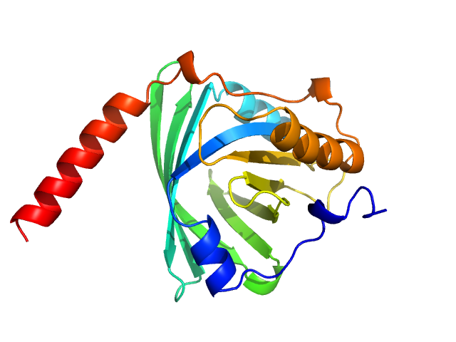
Search

 
Advanced search  

39 hits found for FNU Ashish

SASDMC2 – SWAXS data from Lysozyme Solution [Protein concentration 5 mg/ml] at 10 °C

Lysozyme C experimental SAS data
PDB (PROTEIN DATA BANK) model
Sample: Lysozyme C monomer, 14 kDa Gallus gallus protein
Buffer: 40 mM NaOAc pH 3.8, 150 mM NaCl, pH: 3.8
Experiment: SAXS data collected at X9A, National Synchrotron Light Source (NSLS) on 2014 May 2
Visualizing how inclusion of higher reciprocal space in SWAXS data analysis improves shape restoration of biomolecules: case of lysozyme. J Biomol Struct Dyn :1-15 (2021)
Ashish
RgGuinier 1.3 nm
Dmax 4.2 nm

SASDMD2 – SWAXS data from Lysozyme Solution [Protein concentration 2.5 mg/ml] at 10 °C

Lysozyme C experimental SAS data
PDB (PROTEIN DATA BANK) model
Sample: Lysozyme C monomer, 14 kDa Gallus gallus protein
Buffer: 40 mM NaOAc pH 3.8, 150 mM NaCl, pH: 3.8
Experiment: SAXS data collected at X9A, National Synchrotron Light Source (NSLS) on 2014 May 2
Visualizing how inclusion of higher reciprocal space in SWAXS data analysis improves shape restoration of biomolecules: case of lysozyme. J Biomol Struct Dyn :1-15 (2021)
Ashish
RgGuinier 1.4 nm
Dmax 4.3 nm

SASDME2 – SWAXS data from Lysozyme Solution [Protein concentration 1.8 mg/ml] at 10 °C

Lysozyme C experimental SAS data
PDB (PROTEIN DATA BANK) model
Sample: Lysozyme C monomer, 14 kDa Gallus gallus protein
Buffer: 40 mM NaOAc pH 3.8, 150 mM NaCl, pH: 3.8
Experiment: SAXS data collected at X9A, National Synchrotron Light Source (NSLS) on 2014 May 2
Visualizing how inclusion of higher reciprocal space in SWAXS data analysis improves shape restoration of biomolecules: case of lysozyme. J Biomol Struct Dyn :1-15 (2021)
Ashish
RgGuinier 1.4 nm
Dmax 4.5 nm

SASDMF2 – SWAXS data from Lysozyme Solution [Protein concentration 0.9 mg/ml] at 10 °C

Lysozyme C experimental SAS data
PDB (PROTEIN DATA BANK) model
Sample: Lysozyme C monomer, 14 kDa Gallus gallus protein
Buffer: 40 mM NaOAc pH 3.8, 150 mM NaCl, pH: 3.8
Experiment: SAXS data collected at X9A, National Synchrotron Light Source (NSLS) on 2014 May 2
Visualizing how inclusion of higher reciprocal space in SWAXS data analysis improves shape restoration of biomolecules: case of lysozyme. J Biomol Struct Dyn :1-15 (2021)
Ashish
RgGuinier 1.4 nm
Dmax 5.0 nm

SASDMG2 – SWAXS data from Lysozyme Solution [Protein concentration 0.4 mg/ml] at 10 °C

Lysozyme C experimental SAS data
PDB (PROTEIN DATA BANK) model
Sample: Lysozyme C monomer, 14 kDa Gallus gallus protein
Buffer: 40 mM NaOAc pH 3.8, 150 mM NaCl, pH: 3.8
Experiment: SAXS data collected at X9A, National Synchrotron Light Source (NSLS) on 2014 May 2
Visualizing how inclusion of higher reciprocal space in SWAXS data analysis improves shape restoration of biomolecules: case of lysozyme. J Biomol Struct Dyn :1-15 (2021)
Ashish
RgGuinier 1.5 nm
Dmax 4.6 nm

SASDMH2 – SAXS Data of Lysozyme (Average of 0.9, 1.8, 2.5 and 5 mg/ml) Analyzed in the q range of 0.08-2.75 1/nm

Lysozyme C experimental SAS data
DAMMIF model
Sample: Lysozyme C monomer, 14 kDa Gallus gallus protein
Buffer: 40 mM NaOAc pH 3.8, 150 mM NaCl, pH: 3.8
Experiment: SAXS data collected at X9A, National Synchrotron Light Source (NSLS) on 2014 May 2
Visualizing how inclusion of higher reciprocal space in SWAXS data analysis improves shape restoration of biomolecules: case of lysozyme. J Biomol Struct Dyn :1-15 (2021)
Ashish
RgGuinier 1.4 nm
Dmax 4.2 nm

SASDMJ2 – SAXS Data of Lysozyme (Average of 0.9, 1.8, 2.5 and 5 mg/ml) Analyzed in the q range of 0.08-4.95 1/nm

Lysozyme C experimental SAS data
DAMMIF model
Sample: Lysozyme C monomer, 14 kDa Gallus gallus protein
Buffer: 40 mM NaOAc pH 3.8, 150 mM NaCl, pH: 3.8
Experiment: SAXS data collected at X9A, National Synchrotron Light Source (NSLS) on 2014 May 2
Visualizing how inclusion of higher reciprocal space in SWAXS data analysis improves shape restoration of biomolecules: case of lysozyme. J Biomol Struct Dyn :1-15 (2021)
Ashish
RgGuinier 1.4 nm
Dmax 4.2 nm

SASDMK2 – SWAXS Data of Lysozyme (Average of 0.9, 1.8, 2.5 and 5 mg/ml) Analyzed in the q range of 0.08-9.9 1/nm

Lysozyme C experimental SAS data
DAMMIF model
Sample: Lysozyme C monomer, 14 kDa Gallus gallus protein
Buffer: 40 mM NaOAc pH 3.8, 150 mM NaCl, pH: 3.8
Experiment: SAXS data collected at X9A, National Synchrotron Light Source (NSLS) on 2014 May 2
Visualizing how inclusion of higher reciprocal space in SWAXS data analysis improves shape restoration of biomolecules: case of lysozyme. J Biomol Struct Dyn :1-15 (2021)
Ashish
RgGuinier 1.4 nm
Dmax 4.2 nm

SASDML2 – SWAXS Data of Lysozyme (Average of 0.9, 1.8, 2.5 and 5 mg/ml) Analyzed in the q range of 0.08-17.2 1/nm

Lysozyme C experimental SAS data
DAMMIF model
Sample: Lysozyme C monomer, 14 kDa Gallus gallus protein
Buffer: 40 mM NaOAc pH 3.8, 150 mM NaCl, pH: 3.8
Experiment: SAXS data collected at X9A, National Synchrotron Light Source (NSLS) on 2014 May 2
Visualizing how inclusion of higher reciprocal space in SWAXS data analysis improves shape restoration of biomolecules: case of lysozyme. J Biomol Struct Dyn :1-15 (2021)
Ashish
RgGuinier 1.4 nm
Dmax 4.2 nm

SASDUX3 – F-actin in F-buffer at an actin concentration of 2 mg/mL

Actin, cytoplasmic 1 experimental SAS data
PYMOL model
Sample: Actin, cytoplasmic 1 18-mer, 751 kDa Gallus gallus protein
Buffer: 50 mM KCl, 50 mM Tris-HCl, pH 8.0, 5 mM MgCl2, 1 mM ATP, 0.1% 2-mercaptoethanol, pH: 8
Experiment: SAXS data collected at EMBL P12, PETRA III on 2013 Aug 31
VISUALIZING THE NUCLEATING AND CAPPED STATES OF F-ACTIN BY Ca^2+-ACTIVATED GELSOLIN
FNU Ashish
RgGuinier 13.4 nm
Dmax 48.0 nm

SASDUY3 – Calcium activated full-length gelsolin and the F-form of actin at a 1:32 molar ratio in F-actin buffer

GelsolinActin, cytoplasmic 1 experimental SAS data
Gelsolin Actin, cytoplasmic 1 Kratky plot
Sample: Gelsolin monomer, 84 kDa Homo sapiens protein
Actin, cytoplasmic 1 18-mer, 751 kDa Gallus gallus protein
Buffer: 50 mM KCl, 50 mM Tris-HCl, pH 8.0, 5 mM MgCl2, 1 mM ATP, 0.1% 2-mercaptoethanol, pH: 8
Experiment: SAXS data collected at EMBL P12, PETRA III on 2013 Aug 31
VISUALIZING THE NUCLEATING AND CAPPED STATES OF F-ACTIN BY Ca^2+-ACTIVATED GELSOLIN
FNU Ashish
RgGuinier 12.2 nm

SASDUZ3 – Calcium activated full-length gelsolin and the F-form of actin at a 1:16 molar ratio in F-actin buffer

GelsolinActin, cytoplasmic 1 experimental SAS data
Gelsolin Actin, cytoplasmic 1 Kratky plot
Sample: Gelsolin monomer, 84 kDa Homo sapiens protein
Actin, cytoplasmic 1 18-mer, 751 kDa Gallus gallus protein
Buffer: 50 mM KCl, 50 mM Tris-HCl, pH 8.0, 5 mM MgCl2, 1 mM ATP, 0.1% 2-mercaptoethanol, pH: 8
Experiment: SAXS data collected at EMBL P12, PETRA III on 2013 Aug 31
VISUALIZING THE NUCLEATING AND CAPPED STATES OF F-ACTIN BY Ca^2+-ACTIVATED GELSOLIN
FNU Ashish
RgGuinier 14.5 nm

SASDU24 – Calcium activated full-length gelsolin and the F-form of actin at a 1:8 molar ratio in F-actin buffer

GelsolinActin, cytoplasmic 1 experimental SAS data
Gelsolin Actin, cytoplasmic 1 Kratky plot
Sample: Gelsolin monomer, 84 kDa Homo sapiens protein
Actin, cytoplasmic 1 18-mer, 751 kDa Gallus gallus protein
Buffer: 50 mM KCl, 50 mM Tris-HCl, pH 8.0, 5 mM MgCl2, 1 mM ATP, 0.1% 2-mercaptoethanol, pH: 8
Experiment: SAXS data collected at EMBL P12, PETRA III on 2013 Aug 31
VISUALIZING THE NUCLEATING AND CAPPED STATES OF F-ACTIN BY Ca^2+-ACTIVATED GELSOLIN
FNU Ashish
RgGuinier 13.6 nm

SASDU34 – Calcium activated full-length gelsolin and the F-form of actin at a 1:4 molar ratio in F-actin buffer

GelsolinActin, cytoplasmic 1 experimental SAS data
Gelsolin Actin, cytoplasmic 1 Kratky plot
Sample: Gelsolin monomer, 84 kDa Homo sapiens protein
Actin, cytoplasmic 1 18-mer, 751 kDa Gallus gallus protein
Buffer: 50 mM KCl, 50 mM Tris-HCl, pH 8.0, 5 mM MgCl2, 1 mM ATP, 0.1% 2-mercaptoethanol, pH: 8
Experiment: SAXS data collected at EMBL P12, PETRA III on 2013 Aug 31
VISUALIZING THE NUCLEATING AND CAPPED STATES OF F-ACTIN BY Ca^2+-ACTIVATED GELSOLIN
FNU Ashish
RgGuinier 8.8 nm

SASDU44 – Calcium activated full-length gelsolin and the F-form of actin at a 1:2 molar ratio in F-actin buffer

GelsolinActin, cytoplasmic 1 experimental SAS data
Gelsolin Actin, cytoplasmic 1 Kratky plot
Sample: Gelsolin monomer, 84 kDa Homo sapiens protein
Actin, cytoplasmic 1 18-mer, 751 kDa Gallus gallus protein
Buffer: 50 mM KCl, 50 mM Tris-HCl, pH 8.0, 5 mM MgCl2, 1 mM ATP, 0.1% 2-mercaptoethanol, pH: 8
Experiment: SAXS data collected at EMBL P12, PETRA III on 2013 Aug 31
VISUALIZING THE NUCLEATING AND CAPPED STATES OF F-ACTIN BY Ca^2+-ACTIVATED GELSOLIN
FNU Ashish
RgGuinier 7.3 nm

SASDU54 – Calcium activated full-length gelsolin and the F-form of actin at a 1:1 molar ratio in F-actin buffer

GelsolinActin, cytoplasmic 1 experimental SAS data
Gelsolin Actin, cytoplasmic 1 Kratky plot
Sample: Gelsolin monomer, 84 kDa Homo sapiens protein
Actin, cytoplasmic 1 18-mer, 751 kDa Gallus gallus protein
Buffer: 50 mM KCl, 50 mM Tris-HCl, pH 8.0, 5 mM MgCl2, 1 mM ATP, 0.1% 2-mercaptoethanol, pH: 8
Experiment: SAXS data collected at EMBL P12, PETRA III on 2013 Aug 31
VISUALIZING THE NUCLEATING AND CAPPED STATES OF F-ACTIN BY Ca^2+-ACTIVATED GELSOLIN
FNU Ashish
RgGuinier 5.2 nm

SASDU64 – Calcium-gelsolin and the F-form of actin at a 1:2 molar ratio from high to low ionic strength

GelsolinActin, cytoplasmic 1 experimental SAS data
Gelsolin Actin, cytoplasmic 1 Kratky plot
Sample: Gelsolin monomer, 84 kDa Homo sapiens protein
Actin, cytoplasmic 1 monomer, 42 kDa Gallus gallus protein
Buffer: 2 mM Tris-Cl, pH 8.0, 0.2 mM ATP, 1 mM NaN3, 0.1 mM CaCl2, 0.5 mM DTT, pH: 8
Experiment: SAXS data collected at EMBL P12, PETRA III on 2013 Aug 31
VISUALIZING THE NUCLEATING AND CAPPED STATES OF F-ACTIN BY Ca^2+-ACTIVATED GELSOLIN
FNU Ashish
RgGuinier 4.7 nm
Dmax 25.0 nm
VolumePorod 262 nm3

SASDU74 – Calcium-gelsolin and the F-form of actin at a 1:1 molar ratio from high to low ionic strength

GelsolinActin, cytoplasmic 1 experimental SAS data
Gelsolin Actin, cytoplasmic 1 Kratky plot
Sample: Gelsolin monomer, 84 kDa Homo sapiens protein
Actin, cytoplasmic 1 monomer, 42 kDa Gallus gallus protein
Buffer: 2 mM Tris-Cl, pH 8.0, 0.2 mM ATP, 1 mM NaN3, 0.1 mM CaCl2, 0.5 mM DTT, pH: 8
Experiment: SAXS data collected at EMBL P12, PETRA III on 2013 Aug 31
VISUALIZING THE NUCLEATING AND CAPPED STATES OF F-ACTIN BY Ca^2+-ACTIVATED GELSOLIN
FNU Ashish
RgGuinier 5.2 nm
Dmax 25.0 nm
VolumePorod 266 nm3

SASDU84 – Calcium-gelsolin and the G-form of actin at a 1:2 molar ratio in low ionic strength (SEC-SAXS)

GelsolinActin, cytoplasmic 1 experimental SAS data
GASBOR model
Sample: Gelsolin monomer, 84 kDa Homo sapiens protein
Actin, cytoplasmic 1 monomer, 42 kDa Gallus gallus protein
Buffer: 2 mM Tris-Cl, pH 8.0, 0.2 mM ATP, 1 mM NaN3, 0.1 mM CaCl2, 0.5 mM DTT, pH: 8
Experiment: SAXS data collected at EMBL P12, PETRA III on 2013 Aug 31
VISUALIZING THE NUCLEATING AND CAPPED STATES OF F-ACTIN BY Ca^2+-ACTIVATED GELSOLIN
FNU Ashish
RgGuinier 4.4 nm
Dmax 25.0 nm
VolumePorod 241 nm3

SASDU94 – Calcium-gelsolin and the G-form of actin at a 1:1 molar ratio in low ionic strength (SEC-SAXS)

GelsolinActin, cytoplasmic 1 experimental SAS data
GASBOR model
Sample: Gelsolin monomer, 84 kDa Homo sapiens protein
Actin, cytoplasmic 1 monomer, 42 kDa Gallus gallus protein
Buffer: 2 mM Tris-Cl, pH 8.0, 0.2 mM ATP, 1 mM NaN3, 0.1 mM CaCl2, 0.5 mM DTT, pH: 8
Experiment: SAXS data collected at EMBL P12, PETRA III on 2013 Sep 1
VISUALIZING THE NUCLEATING AND CAPPED STATES OF F-ACTIN BY Ca^2+-ACTIVATED GELSOLIN
FNU Ashish
RgGuinier 4.6 nm
Dmax 25.0 nm
VolumePorod 243 nm3

SASDPG4 – Alpha-1-acid glycoprotein at 283 K

Alpha-1-acid glycoprotein 1 experimental SAS data
PYMOL model
Sample: Alpha-1-acid glycoprotein 1 monomer, 22 kDa Homo sapiens protein
Buffer: phosphate buffered saline, pH: 7.4
Experiment: SAXS data collected at Anton Paar SAXSpace, CSIR - Institute of Microbial Technology (IMTech) on 2021 Jan 12
SAXS data based glycosylated model of alpha-1-acid glycoprotein
FNU Ashish
RgGuinier 2.6 nm
Dmax 7.5 nm
VolumePorod 83 nm3

SASDPH4 – Alpha-1-glycoprotein at 343 K

Alpha-1-acid glycoprotein 1 experimental SAS data
PYMOL model
Sample: Alpha-1-acid glycoprotein 1 monomer, 22 kDa Homo sapiens protein
Buffer: phosphate buffered saline, pH: 7.4
Experiment: SAXS data collected at Anton Paar SAXSpace, CSIR - Institute of Microbial Technology (IMTech) on 2021 Jan 12
SAXS data based glycosylated model of alpha-1-acid glycoprotein
FNU Ashish
RgGuinier 2.5 nm
Dmax 7.5 nm
VolumePorod 96 nm3

SASDN95 – Truncated Kelch13-R539T mutant

Kelch protein K13 (Truncated Kelch13-R539T, artemisinin-resistant mutation) experimental SAS data
GASBOR model
Sample: Kelch protein K13 (Truncated Kelch13-R539T, artemisinin-resistant mutation) hexamer, 396 kDa Plasmodium falciparum (isolate … protein
Buffer: phosphate buffered saline, pH: 7.4
Experiment: SAXS data collected at Anton Paar SAXSpace, CSIR - Institute of Microbial Technology (IMTech) on 2020 Jul 25
Plasmodium falciparum Kelch13 and its artemisinin-resistant mutants assemble as hexamers in solution: a SAXS data driven modeling study. FEBS J (2022)
...Ashish F, Bhattacharjee S
RgGuinier 6.6 nm
Dmax 20.5 nm
VolumePorod 1600 nm3

SASDNA5 – Truncated Kelch13-C580Y mutant

Kelch protein K13 (Truncated Kelch13-C580Y ) experimental SAS data
GASBOR model
Sample: Kelch protein K13 (Truncated Kelch13-C580Y ) hexamer, 397 kDa Plasmodium falciparum (isolate … protein
Buffer: Phosphate Buffer Saline, pH: 7.4
Experiment: SAXS data collected at Anton Paar SAXSpace, CSIR - Institute of Microbial Technology (IMTech) on 2020 Jul 28
Plasmodium falciparum Kelch13 and its artemisinin-resistant mutants assemble as hexamers in solution: a SAXS data driven modeling study. FEBS J (2022)
...Ashish F, Bhattacharjee S
RgGuinier 6.4 nm
Dmax 18.4 nm
VolumePorod 1800 nm3

SASDNB5 – Truncated Kelch13-A578S mutant

Kelch protein K13 (Truncated Kelch13-A578S) experimental SAS data
GASBOR model
Sample: Kelch protein K13 (Truncated Kelch13-A578S) hexamer, 396 kDa Plasmodium falciparum (isolate … protein
Buffer: Phosphate Buffer Saline, pH: 7.4
Experiment: SAXS data collected at Anton Paar SAXSpace, CSIR - Institute of Microbial Technology (IMTech) on 2020 Jul 29
Plasmodium falciparum Kelch13 and its artemisinin-resistant mutants assemble as hexamers in solution: a SAXS data driven modeling study. FEBS J (2022)
...Ashish F, Bhattacharjee S
RgGuinier 6.1 nm
Dmax 16.8 nm
VolumePorod 1700 nm3

SASDN98 – SvGT under calcium free conditions: Apo state

Glyco_trans_2-like domain-containing protein experimental SAS data
OTHER model
Sample: Glyco_trans_2-like domain-containing protein dimer, 96 kDa Streptomyces sp. M41(2017) protein
Buffer: 20 mM Tris, pH 7.5, 100 mM NaCl, and 2 mM DTT + 1 mM EDTA, pH: 7.5
Experiment: SAXS data collected at Anton Paar SAXSpace, CSIR - Institute of Microbial Technology (IMTech) on 2019 Oct 18
Global shape of SvGT, a metal-dependent bacteriocin modifying S/O-HexNActransferase from actinobacteria: c -terminal dimerization modulates the function of this GT. J Biomol Struct Dyn :1-15 (2023)
...Ashish, Rao A
RgGuinier 4.4 nm
Dmax 17.0 nm
VolumePorod 151 nm3

SASDNA8 – SvGT under calcium free conditions: Mg2+ bound state

Glyco_trans_2-like domain-containing protein experimental SAS data
OTHER model
Sample: Glyco_trans_2-like domain-containing protein dimer, 96 kDa Streptomyces sp. M41(2017) protein
Buffer: 20 mM Tris, pH 7.5, 100 mM NaCl, 2 mM DTT, 1 mM EDTA, 2 mM MgCl2, pH: 7.5
Experiment: SAXS data collected at Anton Paar SAXSpace, CSIR - Institute of Microbial Technology (IMTech) on 2019 Oct 18
Global shape of SvGT, a metal-dependent bacteriocin modifying S/O-HexNActransferase from actinobacteria: c -terminal dimerization modulates the function of this GT. J Biomol Struct Dyn :1-15 (2023)
...Ashish, Rao A
RgGuinier 5.8 nm
Dmax 17.9 nm
VolumePorod 111 nm3

SASDLS8 – Complex of Mtb GntR and Aptamer 1 [Rv0792c and Rv0792c_1]

Complex of Rv0792c and Rv0792c_1 experimental SAS data
OTHER model
Sample: Complex of Rv0792c and Rv0792c_1 monomer, 74 kDa
Buffer: 25 mM HEPES Buffer; 400 mM NaCl,, pH: 7.2
Experiment: SAXS data collected at Anton Paar SAXSpace, CSIR - Institute of Microbial Technology (IMTech) on 2019 Oct 31
Structural and Functional Characterization of Rv0792c from Mycobacterium tuberculosis: Identifying Small Molecule Inhibitor against HutC Protein. Microbiol Spectr :e0197322 (2022)
...Ashish, Sharma TK, Singh R
RgGuinier 3.6 nm
Dmax 12.0 nm

SASDLT8 – Complex of Mtb GntR and Aptamer 2 [Rv0792c and Rv0792c_2]

Complex of Rv0792c and Rv0792c_2 experimental SAS data
OTHER model
Sample: Complex of Rv0792c and Rv0792c_2 monomer, 46 kDa
Buffer: 25 mM HEPES Buffer; 400 mM NaCl,, pH: 7.2
Experiment: SAXS data collected at Anton Paar SAXSpace, CSIR - Institute of Microbial Technology (IMTech) on 2019 Oct 31
Structural and Functional Characterization of Rv0792c from Mycobacterium tuberculosis: Identifying Small Molecule Inhibitor against HutC Protein. Microbiol Spectr :e0197322 (2022)
...Ashish, Sharma TK, Singh R
RgGuinier 3.1 nm
Dmax 9.8 nm

SASDLU8 – Complex of Mtb GntR and Aptamer 5 [Rv0792c and Rv0792c_5]

Complex of GntR protein: Aptamer5 experimental SAS data
OTHER model
Sample: Complex of GntR protein: Aptamer5 monomer, 78 kDa
Buffer: 25 mM HEPES Buffer; 400 mM NaCl,, pH: 7.2
Experiment: SAXS data collected at Anton Paar SAXSpace, CSIR - Institute of Microbial Technology (IMTech) on 2019 Oct 31
Structural and Functional Characterization of Rv0792c from Mycobacterium tuberculosis: Identifying Small Molecule Inhibitor against HutC Protein. Microbiol Spectr :e0197322 (2022)
...Ashish, Sharma TK, Singh R
RgGuinier 3.4 nm
Dmax 12.0 nm

SASDLV8 – SS DNA Aptamer 5 which binds Mtb GntR Homolog [Rv0792c_5]

SSDNA Aptamer from SELEX against Mtb Rv0792c experimental SAS data
OTHER model
Sample: SSDNA Aptamer from SELEX against Mtb Rv0792c monomer, 14 kDa unidentified DNA
Buffer: 25 mM HEPES Buffer; 400 mM NaCl,, pH: 7.2
Experiment: SAXS data collected at Anton Paar SAXSpace, CSIR - Institute of Microbial Technology (IMTech) on 2019 Oct 21
Structural and Functional Characterization of Rv0792c from Mycobacterium tuberculosis: Identifying Small Molecule Inhibitor against HutC Protein. Microbiol Spectr :e0197322 (2022)
...Ashish, Sharma TK, Singh R
RgGuinier 1.8 nm
Dmax 7.1 nm

SASDLW8 – SS DNA Aptamer 2 which binds Mtb GntR Homolog [Rv0792c_2]

SSDNA Aptamer from SELEX against Mtb Rv0792c experimental SAS data
OTHER model
Sample: SSDNA Aptamer from SELEX against Mtb Rv0792c monomer, 13 kDa unidentified DNA
Buffer: 25 mM HEPES Buffer; 400 mM NaCl,, pH: 7.2
Experiment: SAXS data collected at Anton Paar SAXSpace, CSIR - Institute of Microbial Technology (IMTech) on 2019 Oct 20
Structural and Functional Characterization of Rv0792c from Mycobacterium tuberculosis: Identifying Small Molecule Inhibitor against HutC Protein. Microbiol Spectr :e0197322 (2022)
...Ashish, Sharma TK, Singh R
RgGuinier 1.9 nm
Dmax 7.6 nm

SASDLX8 – SS DNA Aptamer 1 which binds Mtb GntR Homolog [Rv0792c_1]

SSDNA Aptamer from SELEX against Mtb Rv0792c experimental SAS data
CHIMERA model
Sample: SSDNA Aptamer from SELEX against Mtb Rv0792c monomer, 13 kDa unidentified DNA
Buffer: 25 mM HEPES Buffer; 400 mM NaCl,, pH: 7.2
Experiment: SAXS data collected at Anton Paar SAXSpace, CSIR - Institute of Microbial Technology (IMTech) on 2019 Oct 20
Structural and Functional Characterization of Rv0792c from Mycobacterium tuberculosis: Identifying Small Molecule Inhibitor against HutC Protein. Microbiol Spectr :e0197322 (2022)
...Ashish, Sharma TK, Singh R
RgGuinier 2.1 nm
Dmax 8.6 nm

SASDLY8 – Mycobacterium tuberculosis Rv0792c, a GntR homolog

Recombinant Mycobacterium tuberculosis H37Rv experimental SAS data
OTHER model
Sample: Recombinant Mycobacterium tuberculosis H37Rv dimer, 65 kDa Mycobacterium tuberculosis H37Rv protein
Buffer: 25 mM HEPES Buffer; 400 mM NaCl,, pH: 7.2
Experiment: SAXS data collected at Anton Paar SAXSpace, CSIR - Institute of Microbial Technology (IMTech) on 2019 Oct 2
Structural and Functional Characterization of Rv0792c from Mycobacterium tuberculosis: Identifying Small Molecule Inhibitor against HutC Protein. Microbiol Spectr :e0197322 (2022)
...Ashish, Sharma TK, Singh R
RgGuinier 3.2 nm
Dmax 12.0 nm

SASDLZ8 – SS DNA Aptamer 1 which binds Mtb GntR Homolog [Rv0792c_1]

SSDNA Aptamer from SELEX against Mtb Rv0792c experimental SAS data
DAMMIN model
Sample: SSDNA Aptamer from SELEX against Mtb Rv0792c monomer, 13 kDa unidentified DNA
Buffer: 25 mM HEPES Buffer; 400 mM NaCl,, pH: 7.2
Experiment: SAXS data collected at Anton Paar SAXSpace, CSIR - Institute of Microbial Technology (IMTech) on 2019 Oct 20
Structural and Functional Characterization of Rv0792c from Mycobacterium tuberculosis: Identifying Small Molecule Inhibitor against HutC Protein. Microbiol Spectr :e0197322 (2022)
...Ashish, Sharma TK, Singh R
RgGuinier 2.1 nm
Dmax 8.6 nm

SASDL29 – SAXS data from RW4-MPC at 1 mg/ml at 283 K

Metapyrocatechase experimental SAS data
MODELLER model
Sample: Metapyrocatechase tetramer, 139 kDa Novosphingobium sp. AAP93 protein
Buffer: 20 mM Tris-HCl 250 mM NaCl, pH 8.0, pH: 8
Experiment: SAXS data collected at Anton Paar SAXSpace, CSIR - Institute of Microbial Technology (IMTech) on 2019 Dec 20
Shape-function of a novel metapyrocatechase, RW4-MPC: Metagenomics to SAXS data based insight into deciphering regulators of function. Int J Biol Macromol 188:1012-1024 (2021)
...Ashish, Pinnaka AK
RgGuinier 3.7 nm
Dmax 10.2 nm

SASDNG9 – GIFT based desmeared and regularized SAXS profile on ADP-ribosylation factor-like protein 15 (apo conditions)

ADP-ribosylation factor-like protein 15 experimental SAS data
GASBOR model
Sample: ADP-ribosylation factor-like protein 15 monomer, 22 kDa Homo sapiens protein
Buffer: 20 mM Tris, 100 mM NaCl, 1 mM PMSF, 10% glycerol, 1 mM DTT, pH: 8
Experiment: SAXS data collected at Anton Paar SAXSpace, CSIR - Institute of Microbial Technology (IMTech) on 2018 Jul 10
ARL15, a GTPase implicated in rheumatoid arthritis, potentially repositions its truncated N-terminus as a function of guanine nucleotide binding International Journal of Biological Macromolecules 254:127898 (2024)
...Ashish F, Kundu S
RgGuinier 1.8 nm
Dmax 6.1 nm
VolumePorod 33 nm3

SASDNH9 – GIFT based desmeared and regularized SAXS profile on ADP-ribosylation factor-like protein 15 with GDP

ADP-ribosylation factor-like protein 15 experimental SAS data
GASBOR model
Sample: ADP-ribosylation factor-like protein 15 monomer, 22 kDa Homo sapiens protein
Buffer: 20 mM Tris, 100 mM NaCl, 1 mM PMSF, 10% glycerol, 1 mM DTT, pH: 8
Experiment: SAXS data collected at Anton Paar SAXSpace, CSIR - Institute of Microbial Technology (IMTech) on 2018 Jul 10
ARL15, a GTPase implicated in rheumatoid arthritis, potentially repositions its truncated N-terminus as a function of guanine nucleotide binding International Journal of Biological Macromolecules 254:127898 (2024)
...Ashish F, Kundu S
RgGuinier 2.0 nm
Dmax 7.6 nm
VolumePorod 38 nm3

SASDNJ9 – GIFT based desmeared and regularized SAXS profile on ADP-ribosylation factor-like protein 15 with GTP

ADP-ribosylation factor-like protein 15 experimental SAS data
GASBOR model
Sample: ADP-ribosylation factor-like protein 15 monomer, 22 kDa Homo sapiens protein
Buffer: 20 mM Tris, 100 mM NaCl, 1 mM PMSF, 10% glycerol, 1 mM DTT, pH: 8
Experiment: SAXS data collected at Anton Paar SAXSpace, CSIR - Institute of Microbial Technology (IMTech) on 2018 Jul 10
ARL15, a GTPase implicated in rheumatoid arthritis, potentially repositions its truncated N-terminus as a function of guanine nucleotide binding International Journal of Biological Macromolecules 254:127898 (2024)
...Ashish F, Kundu S
RgGuinier 2.0 nm
Dmax 7.7 nm
VolumePorod 38 nm3