|
|
|
|
|
| Sample: |
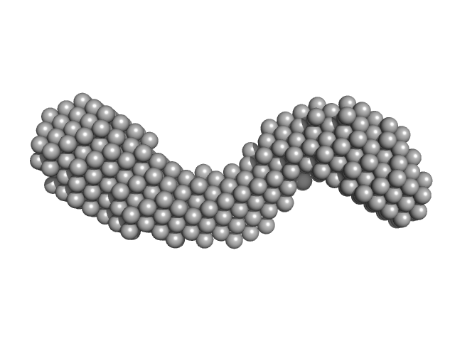
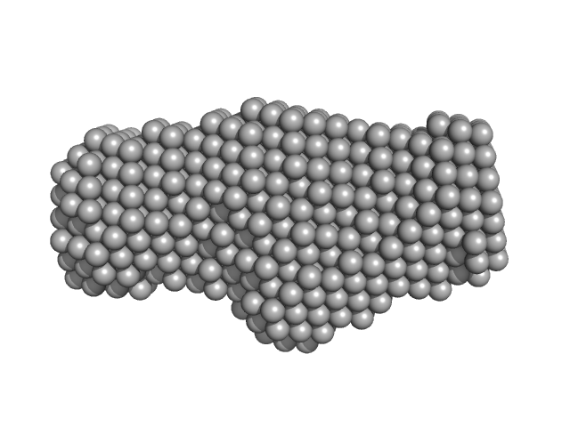
E3 ubiquitin-protein ligase LRSAM1 monomer, 84 kDa Homo sapiens protein
|
| Buffer: |
25 mM Tris-HCl pH 8.0, 150 mM NaCl, 1 mM TCEP, pH: 8 |
| Experiment: |
SAXS
data collected at BL-10C, Photon Factory (PF), High Energy Accelerator Research Organization (KEK) on 2019 Dec 7
|
Leucine Rich Repeat And Sterile Alpha Motif Containing 1 (LRSAM1)
Si Hoon Park
|
| RgGuinier |
6.1 |
nm |
| Dmax |
19.4 |
nm |
| VolumePorod |
250 |
nm3 |
|
|
|
|
|
|
|
| Sample: |
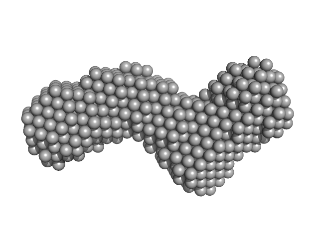
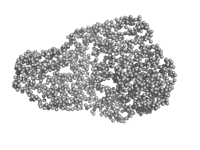
E3 ubiquitin-protein ligase LRSAM1 - ΔPTAP/PSAP-RING monomer, 74 kDa Homo sapiens protein
|
| Buffer: |
50 mM Tris-HCl pH 8.0, 300 mM NaCl, 1 mM TCEP, 5% w/v glycerol, pH: 8 |
| Experiment: |
SAXS
data collected at 4C, Pohang Accelerator Laboratory on 2017 Oct 24
|
Leucine Rich Repeat And Sterile Alpha Motif Containing 1 (LRSAM1)
Si Hoon Park
|
| RgGuinier |
5.5 |
nm |
| Dmax |
18.0 |
nm |
| VolumePorod |
322 |
nm3 |
|
|
|
|
|
|
|
| Sample: |
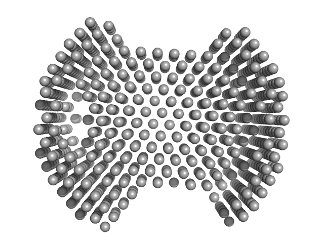
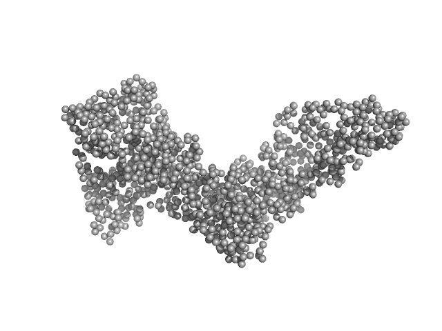
E3 ubiquitin-protein ligase LRSAM1 - LRR containing N-terminal domain monomer, 35 kDa Homo sapiens protein
|
| Buffer: |
50 mM Tris-HCl pH 8.0, 300 mM NaCl, 1 mM TCEP, 5% w/v glycerol, pH: 8 |
| Experiment: |
SAXS
data collected at 4C, Pohang Accelerator Laboratory on 2017 Oct 23
|
Leucine Rich Repeat And Sterile Alpha Motif Containing 1 (LRSAM1)
Si Hoon Park
|
| RgGuinier |
3.4 |
nm |
| Dmax |
8.2 |
nm |
| VolumePorod |
105 |
nm3 |
|
|
|
|
|
|
|
| Sample: |
E3 ubiquitin-protein ligase LRSAM1 - SAM domain monomer, 8 kDa Homo sapiens protein
|
| Buffer: |
50 mM Tris-HCl pH 8.0, 150 mM NaCl, 1 mM TCEP, pH: 8 |
| Experiment: |
SAXS
data collected at 4C, Pohang Accelerator Laboratory on 2019 Apr 4
|
Leucine Rich Repeat And Sterile Alpha Motif Containing 1 (LRSAM1)
Si Hoon Park
|
| RgGuinier |
1.4 |
nm |
| Dmax |
4.3 |
nm |
| VolumePorod |
11 |
nm3 |
|
|
|
|
|
|
|
| Sample: |
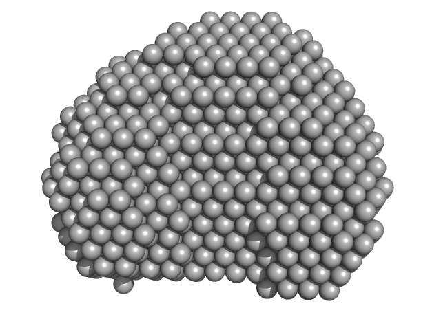
E3 ubiquitin-protein ligase LRSAM1 monomer, 30 kDa Homo sapiens protein
|
| Buffer: |
50 mM Tris-HCl pH 8.0, 150 mM NaCl, 1 mM TCEP, pH: 8 |
| Experiment: |
SAXS
data collected at 4C, Pohang Accelerator Laboratory on 2019 Apr 4
|
Leucine Rich Repeat And Sterile Alpha Motif Containing 1 (LRSAM1)
Si Hoon Park
|
| RgGuinier |
2.5 |
nm |
| Dmax |
8.3 |
nm |
| VolumePorod |
43 |
nm3 |
|
|
|
|
|
|
|
| Sample: |
Gelsolin monomer, 84 kDa Homo sapiens protein
Actin, cytoplasmic 1 monomer, 42 kDa Gallus gallus protein
|
| Buffer: |
2 mM Tris-Cl, pH 8.0, 0.2 mM ATP, 1 mM NaN3, 0.1 mM CaCl2, 0.5 mM DTT, pH: 8 |
| Experiment: |
SAXS
data collected at EMBL P12, PETRA III on 2013 Aug 31
|
Visualizing the nucleating and capped states of f-actin by Ca(2+)-gelsolin: Saxs data based structures of binary and ternary complexes.
Int J Biol Macromol :134556 (2024)
Sagar A, Peddada N, Choudhary V, Mir Y, Garg R, Ashish
|
| RgGuinier |
4.4 |
nm |
| Dmax |
25.0 |
nm |
| VolumePorod |
241 |
nm3 |
|
|
|
|
|
|
|
| Sample: |
Gelsolin monomer, 84 kDa Homo sapiens protein
Actin, cytoplasmic 1 monomer, 42 kDa Gallus gallus protein
|
| Buffer: |
2 mM Tris-Cl, pH 8.0, 0.2 mM ATP, 1 mM NaN3, 0.1 mM CaCl2, 0.5 mM DTT, pH: 8 |
| Experiment: |
SAXS
data collected at EMBL P12, PETRA III on 2013 Sep 1
|
Visualizing the nucleating and capped states of f-actin by Ca(2+)-gelsolin: Saxs data based structures of binary and ternary complexes.
Int J Biol Macromol :134556 (2024)
Sagar A, Peddada N, Choudhary V, Mir Y, Garg R, Ashish
|
| RgGuinier |
4.6 |
nm |
| Dmax |
25.0 |
nm |
| VolumePorod |
243 |
nm3 |
|
|
|
|
|
|
|
| Sample: |
Ubiquitin carboxyl-terminal hydrolase 8 monomer, 127 kDa Homo sapiens protein
|
| Buffer: |
25 mM Tris, 150 mM NaCl, pH: 7.5 |
| Experiment: |
SAXS
data collected at BioCAT 18ID, Advanced Photon Source (APS), Argonne National Laboratory on 2022 Jun 18
|
Autoinhibition of ubiquitin-specific protease 8: insights into domain interactions and mechanisms of regulation
Journal of Biological Chemistry :107727 (2024)
Caba C, Black M, Liu Y, DaDalt A, Mallare J, Fan L, Harding R, Wang Y, Vacratsis P, Huang R, Zhuang Z, Tong Y
|
| RgGuinier |
8.4 |
nm |
| Dmax |
31.8 |
nm |
| VolumePorod |
327 |
nm3 |
|
|
|
|
|
|
![OTHER [STATIC IMAGE] model](/media/pdb_file/SASDV54_fit1_model1.png)
|
| Sample: |
Phage antirepressor protein Cro dimer, 24 kDa Escherichia coli O157:H7 protein
IR1 22 base pair dsDNA monomer, 14 kDa DNA
|
| Buffer: |
20 mM sodium acetate, 150 mM NaCl, 1 mM TCEP, pH: 5.5 |
| Experiment: |
SAXS
data collected at BM29, ESRF on 2021 Apr 13
|
Structural-function relationship of YdaS, a Cro-type repressor in the cryptic prophage CP-933P from Escherichia coli O157:H7
Marusa Prolic Kalinsek
|
| RgGuinier |
2.8 |
nm |
| Dmax |
10.1 |
nm |
| VolumePorod |
53 |
nm3 |
|
|
|
|
|
|
|
| Sample: |
Z-DNA-binding protein 1 monomer, 18 kDa Homo sapiens protein
|
| Buffer: |
20 mM Tris, 100 mM NaCl, pH: 7.5 |
| Experiment: |
SAXS
data collected at BL19U2, Shanghai Synchrotron Radiation Facility (SSRF) on 2018 Mar 23
|
ZBP1 condensate formation synergizes Z-NAs recognition and signal transduction.
Cell Death Dis 15(7):487 (2024)
Xie F, Wu D, Huang J, Liu X, Shen Y, Huang J, Su Z, Li J
|
| RgGuinier |
2.1 |
nm |
| Dmax |
9.0 |
nm |
| VolumePorod |
19 |
nm3 |
|
|


